
Prentice Hall Biology (California)
1st Edition
Kenneth R. Miller, Levine
ISBN: 9780132013529
Textbook solutions

All Solutions

Page 939: Chapter Assessment

Exercise 1
Step 1
1 of 2
Bones are composed of solid living tissues and protein fibers and calcium salt deposits. The tough outer layer is called periosteum.
Result
2 of 2
C. periosteum
Exercise 2
Step 1
1 of 2
Just below the periosteum is the compact bone which contains a network of tubes called Haversian canals.
Result
2 of 2
c. Haversian canals
Exercise 3
Step 1
1 of 2
The process of bone formation is called ossification. In ossification, the bone tissues are formed to replace the cartilage.
Result
2 of 2
A. ossification
Exercise 4
Step 1
1 of 2
Ligaments are strips of tough yet flexible connective tissues that connect two bones together.
Result
2 of 2
D. ligaments
Exercise 5
Step 1
1 of 2
A fluid-filled sac found between the joints in the body is called bursae. These sacs are filled with synovial fluid that helps provide cushion and reduce friction between bones.
Result
2 of 2
A. bursae
Exercise 6
Step 1
1 of 2
The ball-and-socket joints permit rotating and swinging movements.
Result
2 of 2
B. ball-and-socket joints
Exercise 7
Step 1
1 of 2
Figure A shows a skeletal muscle.
Figure B shows a cardiac muscle.
Figure C shows a smooth muscle.
Figure D shows a Haversian canal.

Therefore, figure C is the correct answer.
Result
2 of 2
Figure C (smooth muscle)
Exercise 8
Step 1
1 of 2
During muscle contraction, a cross-bridge is created between two proteins, which are the myosin (thick) filaments and the actin (thin) filaments.
Result
2 of 2
B. actin and myosin
Exercise 9
Step 1
1 of 2
The neuromuscular junction serves as the area of communication or contact between a motor neuron and a skeletal muscle fiber.
Result
2 of 2
D. neuromuscular junction
Exercise 10
Step 1
1 of 2
The outermost covering of the skin is called the epidermis.
Result
2 of 2
C. epidermis
Exercise 11
Result
1 of 1
The skeletal system is made up of connective tissues that include bones, cartilage, tendons, and ligaments.
Exercise 12
Result
1 of 1
Bones are rigid living tissues that provide support and structure to the body. They are composed of living cells, protein fibers, and deposits of calcium salts.
Exercise 13
Step 1
1 of 2
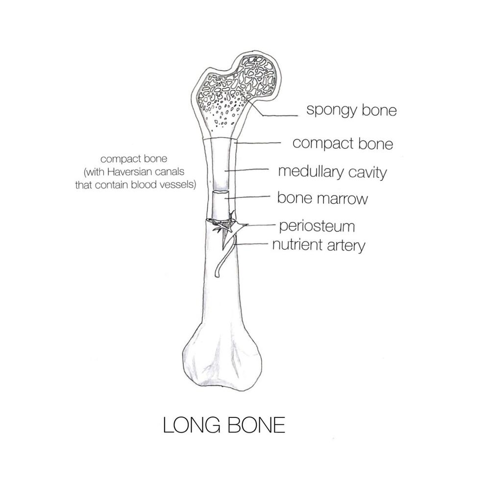
Below is a diagram that shows the structure of a long bone. A long bone contains Haversian canals wherein the blood vessels and nerves are located. These structures carry oxygen and nutrients to the bones.Exercise scan
Result
2 of 2
A long bone contains Haversian canals wherein the blood vessels and nerves are located. These structures carry oxygen and nutrients to the bones. Click to see the diagram that shows the structure of a long bone.
Exercise 14
Result
1 of 1
The spongy bone’s less dense structure allows the near ends of long bones to have an added strength without adding mass. This makes the bones lighter and prevents damage since force is usually applied at the ends of the bones. In addition, the spongy bone contains pores that are filled with blood vessels, which deliver essential nutrients and oxygen to the rest of the bones.
Exercise 15
Result
1 of 1
The red bone marrow generates stem cells that develop into red blood cells, white blood cells, and platelets.
Exercise 16
Step 1
1 of 2
There are three types of muscles found in the human body: s**mooth muscle, skeletal muscle**, and **cardiac muscle**. **Smooth muscle** is characterized as having involuntary, non-striated, uninucleate, and unbranched cells. **Skeletal muscle**, on the other hand, is described as voluntary, striated, multinucleated, and unbranched. Lastly, the **cardiac muscle** is the only muscle that forms our heart. it is composed of involuntary, striated, branched cells, and intercalated discs that connect one cardiac muscle to another.
Step 2
2 of 2

Both **smooth** and **cardiac muscles** are designed well to facilitate the involuntary contraction and movement of the internal organs including the function of the heart to pump blood and the peristalsis motion of the gastrointestinal tract. On the other hand, **skeletal muscles** regulate voluntary movements which can be consciously controlled by ourselves.

Exercise 17
Result
1 of 1
Muscle contraction happens when the muscle proteins slide past each other to produce a movement. This event is called the sliding filament model of muscle contraction. During contraction, a cross-bridge is created between two proteins, which are the myosin (thick) filaments and the actin (thin) filaments. Once the cross-bridge is formed, it changes shape and starts to pull the actin filaments. This results to the actin filaments sliding toward the sarcomere, then it detaches from the cross-bridge.
Exercise 18
Step 1
1 of 2
**Skeletal muscles** function to regulate and control the various voluntary movements of the body. In order to accomplish this function, these muscle tissues should undergo contraction. Contraction is made possible through the connection of muscle tissues to the motor neurons, where the portion called **neuromuscular junction** specifically connects the skeletal muscle cell to the motor neuron.
Step 2
2 of 2
The axon terminals of the motor neuron release a neurotransmitter called **acetylcholine** that diffuses across the synapse, hence creating a signal or impulse in the cell membrane of the muscle cell. This signal will then result in the release of calcium ions in the cell, allowing the interaction of actin and myosin fibers, thus resulting in *muscle contraction*.
Exercise 19
Result
1 of 1
Ligaments bind bones together and stabilize the joints, whereas tendons hold muscles to the bones.
Exercise 20
Result
1 of 1
Since skeletal muscles pull the bones in one direction, they are not able to return to their original position. Because of this, muscles work in opposing pairs–one muscle is bending or flexing (contracting) and the other one is extending (relaxing). For example, when you are bending your elbow, the biceps contract, while the triceps relax.
Exercise 21
Step 1
1 of 3
The **integumentary system** functions as a barrier against the external environment and the body. Moreover, this system also helps in temperature regulation, removal of waste materials, and protection against the UV rays of the sun.
Step 2
2 of 3
One of the most important functions of the integumentary system is **temperature regulation**. The skin connects and interacts with other body systems in order to balance and maintain homeostasis. The skin specifically helps in regulating the body temperature by regulating the blood vessels present in the dermis. During hot weather, the blood vessels in the dermis area dilate to transport the heat from the body to the skin, while during cold weather, the blood vessels constrict conserving the heat in the body.
Step 3
3 of 3
Moreover, the dermis of the skin contains sweat glands that regulate the perspiration in the body. As the sweat is released it will evaporate and the heat and waste materials are also being let go from the body.
Exercise 22
Step 1
1 of 2
The **integumentary system** functions as a barrier against the external environment and the body. Moreover, this system also helps in temperature regulation, removal of waste materials, and protection against the UV rays of the sun. The biggest and major component of the integumentary system is the **skin**.
Step 2
2 of 2
The skin is composed of two layers, the *epidermis* and *dermis*.

1. The **epidermis** is the outer layer of the skin composed of two layers in which the outer layer is comprised of dead skin cells and the inner layer is comprised of proliferating cells. These cells push the older cells to the surface of the epidermis, and as it moves upward the cells tend to disintegrate and die.

2. The **dermis** is the layer beneath the epidermis. This layer of the skin is composed of collagen fibers, blood vessels, nerve endings, follicles, and smooth muscles. The dermis also contains the two important glands of the skin which are the sebaceous gland and sweat gland.

Exercise 23
Step 1
1 of 2
The **epidermis** is the outer layer of the skin composed of two layers in which the out layer is comprised of dead skin cells and the inner layer is comprised of proliferating cells. These cells push the older cells to the surface of the epidermis, and as it moves upward the cells tend to disintegrate and die.
Step 2
2 of 2
The epidermis also contains *melanocytes*, which function to produce and release the pigment **melanin**– a dark brown pigment, that plays an important role in the protection of the skin against UV rays, and also affects skin color.

Although most people have the relative same amount of melanocytes in the skin, the production of the pigment melanin differs, and this result in differences in the skin color among individuals.

Exercise 24
Step 1
1 of 2
The **skin** connects and interacts with other body systems in order to balance and maintain homeostasis. The skin specifically helps in regulating the body temperature by regulating the blood vessels present in the dermis. During hot weather, the blood vessels in the dermis area dilate to transport the heat from the body to the skin, while during cold weather, the blood vessels constrict conserving the heat in the body.

Step 2
2 of 2
Moreover, the **dermis of the skin contains sweat glands that regulate the perspiration in the body**. As the sweat is released it will evaporate and the heat and waste materials are also being let go from the body.
Exercise 25
Result
1 of 1
As the cells divide from the nail root, the new cells push the old nails in the fingers and toes. The old cells become flat and start to harden. The old nail located above the skin is made up of dead cells that are covered in keratin.
Exercise 26
Result
1 of 1
The disks of cartilage between the bones in the spinal column, which are called the intervertebral disks, act as a shock absorber that provides protection to the spine. They give flexibility and mobility to the spinal column.
Exercise 27
Result
1 of 1
A muscle will stop its contraction once the production of a neurotransmitter called acetylcholine stops and an enzyme destroys the remaining acetylcholine. Acetylcholine helps trigger an impulse in the muscle fibers. If an impulse is stopped due to the toxins from the bacteria, the interaction between muscle fibers will not happen; hence, the muscles would not be able to pull the bones and create a movement.
Exercise 28
Step 1
1 of 1
Cartilage is a type of connective tissue that is mostly present during the **embryonic, infant, and toddler stages**. This connective tissue is then replaced by bone structures through the process of ossification as the individual gets older. Therefore, based on the given X-ray figure, the person who has the highest amount of cartilage, and the youngest among them, is the person that has **left-most side hand of the picture**.
Exercise 29
Step 1
1 of 2
**Osteoporosis** is one of the common diseases in the bones. This disease is characterized by having *brittle bones* to the point where the bony structures can easily be broken. Interestingly, this disease is very frequent among women, because women’s bones are relatively thinner and smaller compared to men’s bones
Step 2
2 of 2
This is the reason why most doctors advise older people, especially older women to take calcium supplements to prevent bone weakening. In addition, to this, weight-bearing exercises are also recommended to further strengthen the bones.
Exercise 30
Step 1
1 of 2
If we want to build a robotic arm that replicates the working of a human elbow, then we need to consider three important aspects of the working of the elbow joint.
Step 2
2 of 2
The elbow joint consists of three bones, humerus, ulna, radius, and three types of joints.

1. Joint between the trochlea of humerus and ulna, which is a hinge joint permitting back and forth motion.

2. Joint between capitulum of humerus and radius head, which is also a hinge joint.

3. Upper part of the radius and ulna form a pivot joint. Such a joint allows one bone to rotate over the other bone.

These joints are very essential to replicate the elbow in the robotics arm.

Exercise 31
Step 1
1 of 1
Elbow is the region of a joint at this point two bones come in contact, which is held together by a ligament These bones are attached to muscles by the tendons. Therefore, repeated leaning might have extra pressure either on the ligaments or tendons, leading to an injury to either of them, which may be a possible cause of painful swelling.
Exercise 32
Step 1
1 of 1
Blood vessels that carry oxygen and nutrients to different parts of the body are arteries. Along with these, they also carry blood cells which are essential for the healing process. Especially, white blood cells and platelets are involved in the healing process. Since ligaments are poorly supplied by blood vessels, injury to ligaments is painful and takes a much longer time to heal. As it is clear that lack of blood supply restricts the supply of nutrients, oxygen, white blood cells, and platelets reducing the time of healing of a ligament injury.
Exercise 33
Step 1
1 of 2
Effects of over-exercising:- The most apparent effect of over-exercising is dehydration due to inadequate fluid intake. Over-exercising with dieting can slow down the metabolism process and loss of appetite.
Step 2
2 of 2
Other effects of over-exercising are:
1) Insomnia, depression, fatigue, headache
2) Physical effects include – muscular and skeletal injuries i.e., fractural bones arthritis, etc,
3) Also linked to cellular mutations and cancer as over-exercising can lead to the release of excessive free radicals.
4) It can lead to problems related to heart

In women, it may lead to amenorrhea.

Exercise 34
Step 1
1 of 2
Skin generally protects the body from ultraviolet (UV) radiations emitted by the sun. Too much exposure to UV rays can cause skin cancer. To protect from sunburn, we are advised to wear sunglasses and a hat. Moreover, it is recommended to apply effective sunscreen. Each sunscreen comes with a sun protection factor (SPF), which is applied depending upon the time of exposure to the sun.

Step 2
2 of 2
A company that produces sunscreen having SPF 15 claims to protect from sunburn due to UV rays. Basically, sunscreen with SPF 15 can protect for 4 to 8 hours from sunburn. For that, first, apply sunscreen of SPF 15 and get exposed to rays with minimal UV index level (02), which usually burns skin in 60 minutes. If skin shows sunburn before the prescribed time, the claim of sunscreen is wrong.
Exercise 35
Step 1
1 of 1
Callus develops because of excessive pressure over a certain area, callus often occurs in the foot because the foot supports the body weight while standing and walking. Other reasons for callus development on the foot may be high – heeled shoes, gait abnormalities, highly arched foot, obesity, and excessively long metatarsal bone of the foot.
unlock
Get an explanation on any task
Get unstuck with the help of our AI assistant in seconds
New