Visual system anatomy and Physiology – Flashcards
Unlock all answers in this set
Unlock answersquestion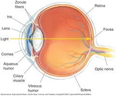
what's Sclera

answer
the outermost layer of the eye and is a tough white fibrous tissue Sclera muscles
question
Pupil
answer
The pupil is an opening that allows light to enter the eye.
question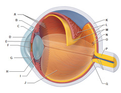
Cornea

answer
A transparent gelatinous structure in front of Iris which allows light in.it is nourished by the aqueous humour
question
The anterior chamber.
answer
The space beteen lens & cornea filled with the aqueous humour
question
Posterior chamber
answer
• Between lens & Iris.
question
Canal of schelm
answer
A canal at junction of cornea and iris through which aqueous humour flows
question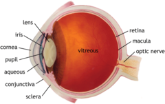
Vitreous humour

answer
The middle of eye , jelly-like Vitreous humor makes up 80% of eye and keeps eyeballs spherical . need constant replacement + has phagocytes which breakdown and remove obstacles/ floaters.
question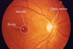
Macula

answer
• The macula is a spot at the back of the eye responsible for central vision.
question
whats at the centre of macula?
answer
fovea
question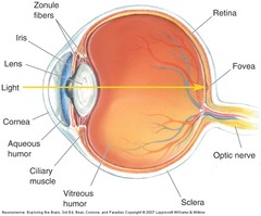
Optic disk

answer
• A region at the back of eye continuous with the retina which is the origin of blood vessels and where the optic nerve axons exit eye
question
why is the optic disk called a blind spot
answer
nophotoreceptors
question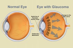
what causes glaucoma

answer
disorder caused by imbalance of aqueous humour due to increased pressure reducing blood supply to the eye thus damaging retina
question
Transduction of light
answer
• Light passes pass through the cornea, lens, and anterior and posterior chambers and reach the photoreceptors (rods and cones) located in the retina • Focusing of images on the photoreceptors depends on refraction (bending) of light rays as they pass through the cornea and the lens
question
photoreceptor types
answer
rods and cones
question
do photoreceptors producs APs?
answer
no,They don't not produce APs. They respond with graded membrane potentials .
question
describe cones
answer
- responsible for daylight, colour and acuity vision -concentrated at fovea- central vision -They have a fast integration time and response and are sensitive to direct axial light due to the cone shape. -least numerous (5 million) -• Cell bodies are located in outer nuclear layer of retina have 3 photopigments with different peak sensitivities ( SML cones)
question
what happens due to loss of cones
answer
decreased visual acuity and colour vision deficits
question
describe rods
answer
- responsible for night vision have more discs to absorb light. Their disks contain lightabsorbing photopigments. -more numerous (100 million) -slow integration -1 photopigment -Cell bodies are located in outer nuclear layer of retina -dominate peripheral retina -contain rhodopsin pigment which is highly sensitive to light allowing night vision its peak sensitivity ~ 496 nm (blue/green)
question
how is colour perceived
answer
Comparison of two areas of equal light intensity reflecting different wavelengths
question
types of cones, colour and max nm activation
answer
S-cone -- short wavelength (430nm max) ---> blue M cone --> medium wavelength (530 nm) --> green L cone---> large walength (560max) ---> red
question
Trichromacy
answer
the condition of possessing three independent channels for conveying color information, derived from the three different cone types
question
describe the inner nuclear layer of the eye
answer
• Contains the cell bodies of amacrine cells, horizontal cells, and bipolar cells which function as interneurons. • Horizontal and bipolar cells process signals from photoreceptors.
question
phototransduction
answer
• The right and left lateral geniculate nuclei, located in the dorsal thalamus, are the major targets of the two optic tracts. • Light ---> eye retina - to photoreceptors • Absorbed by pigments complex (rhodopsin (rods)/ conopsin cones) & cis-retinal ) • Light causes conformational change from cis-RETINAL to trans-retinal • Complex - GDPGTP - activates cGMP phosphodiesterase which Breaks down cGMP tp gmp results in closure of sodium channels • "dark current" prevented - Na+ leaves but cannot enter • Cell hyperpolarises - reduces transmitter released
question
what happens when light hits rod
answer
it turns off via closure of na+ channels
question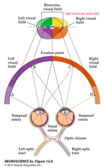
describe the Visual hemifieldfield production

answer
• light from periphery hits the nasal retina and travels contralaterally crossing at the optic chiasm to the LGN • light from the visual field hits the temporal retina and travels ipsilateraly to the LGN,then optic radiation • Objects within binocular field (B) of left hemi-field are imaged by nasal retina of left eye but also temporal retina of right eye • Objects outside of left binocular field (A) imaged by nasal retina of left eye • Fibres from nasal retina cross (decussate) at optic chiasm • NB Left visual hemi-field is viewed by right hemi-sphere and vice versa
question
Visual pathways
answer
• light goes to the retina lateral geniculate nucleus primary visual cortex
question
whats a full visual field (F)
answer
area viewed by both eyes
question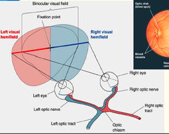
Visual Hemifields

answer
• Full visual field (FVF) is area viewed by both eyes • Left/Right hemi-field divides FVF into two • Binocular VF is area of overlap between left & right hemi-fields
question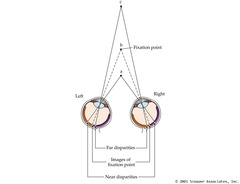
fixation points

answer
• Fixation point (b) - represents focus point for object • Near (a) & far (b) disparities - areas in front and behind where two images are perceived. • Perceptual disparities -interpreted as 'depth' by brain
question
what is interpreted as depth?
answer
perceptual disparities
question
Retinofugal projection
answer
• Pathway of optic nerve to brain • R & L optic nerves to optic chiasm • The chiasm is site of a decussation (crossing) so that the L visual field from both eyes projects to the R cortical hemisphere and R field to L hemisphere. • The nasal part of the retina crosses to contralateral side the temporal part remains ipsilateral • Axons project to R and L Lateral geniculate nucleus (LGN) in R and L thalamus • R and L optic radiations to primary visual cortex (aka, area 17, V1 or striate cortex) of R and L occipital lobes of cerebral cortex
question
Blindsight
answer
a type of vision "which is not dependent on primary areas responsible for processing vision" • "Thought to result from the projections of the optic nerves to subcortical areas, including the superior colliculus and the amygdala" • "Part of our vision that's for orienting and doing rather than for understanding"
question
LGN projections
answer
• Neurones of LGN project to visual cortex via optic radiation
question
LGN target
answer
• Main target is primary visual cortex Also referred to as Brodmann's area 17, the striate cortex or V1 • Lies in occipital lobe • Laminar structure
question
striate cortex
answer
Striate" cortex - striped- has neuronal bodies arranged in layers which can be seen by staining the cortex . named can also be seen in fresh tissue caused by • Due to horizontal pyramidal cell connections in layer III
question
Visual Association Cortex
answer
• Early studies identified Brodmann's areas 17 (V1), 18 & 19 as 'visual' • Actually > 50 % primate cortex subserves vision, V1-V5 • Dorsal stream - motion processing • Ventral stream - visual details, face selective cells
question
RF 'centre' contact with bipolar cell
answer
direct
question
RF 'surround' contact with bipolar cell
answer
via horizontal cell
question
what's a receptive field
answer
area of retina that alters bipolar Vm in response to light
question
describe off centre bipolar cells
answer
-they depolarise in the dark when light hits surround they receive indirect input from receptive field centre the photoreceptor is hyperpolarised and this acts on horizontal cell also hyperpolarises horizontal cell acts on bipolar cells and hyperpolarises them
question
On centre cells
answer
-depolarised when light falls on centre -light on surround hyperpolarises cells • Reduces horizontal cell output • Reduces GABA inhibition of centre • More glutamate released in centre so ON cell hyperpolarises
question
types of ganglion cells
answer
• Magnocellular ganglion cells (M-type) • Parvocellular ganglion cells (P-type): • K ganglion cells (non-M, non-P type)
question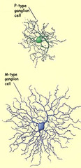
•Magnocellular ganglion cells (M-type)

answer
o 5% of ganglion cell population o larger, more numerous, for scotopic vision & motion detection o respond to combined M & L cones and rods o brightness contrast only
question
• Parvocellular ganglion cells (P-type):
answer
o 90% of ganglion cell population o small, fewer, for colour vision o respond to single cones or groups of same cones o colour opponent cells
question
• K ganglion cells (non-M, non-P type)
answer
o small cell bodies, medium-sized dendritic fields, colour selective o 5% ganglion population
question
Ganglion cell RF's
answer
• Have antagonistic centre-surround receptive fields (RF) • 'On-centre' and 'Off-centre' ganglion cell subtypes • 'On-centre' - depolarize in light, hyperpolarize in dark • 'Off-centre' - hyperpolarize in light, depolarize in dark • Optimal encoding of differences in illumination across RF
question
Off" ganglion cells
answer
• Shadow in 'surround' RF only - hyperpolarisation & decreased firing • As 'edge' moves over 'centre' get depolarization & increased firing • When shadow completely covers 'centre' & 'surround' then firing decreases again • Conclude that optimal stimulus is dark-light border across 'centre' & 'surround' RF i.e. spatial variation
question
What purpose does this centre-surround antagonistic organisation serve?
answer
: It helps the retina find edges in images. Objects are usually distinguished from their background by sudden changes in reflected light
question
Colour Opponency
answer
•Colour perception is based on relative activity of ganglion cells whose RF centres receive inputs from red, green & blue cones • Looking at red box fatigues some of red cones • When we switch our vision to the white box, the green cones (for the red square) are unopposed, so we see green • Same for blue box - blue cones become fatigued so we see yellow when we switch our vision • Ganglion cells - important for providing spatial comparison information about light/dark, red/green, and blue/yellow
question
what type of ganglion cell is Red ON centre green OFF surround
answer
P-cell
question
describe the stimuli of red and green on P cells
answer
• RF - red ON centre; green OFF surround (R+G-) • Most effective stimulus is red light onto RF centre • Response strongly inhibited by green light onto RF surround • Some cells classed as blue ON & yellow OFF (B+Y-)
question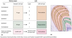
what layers of the LGN do magnocellular g cells target

answer
: layers 1 & 2, inputs
question
what layers of the LGN do parvocellular g cells target
answer
layers 3-6
question
LGN targets: organisation
answer
• Retinotopic projection • Neighbouring locations on the retina project to neighbouring locations on the LGN • The projection pattern is preserved in the LGNV1 projection
question
in what layer of the cortex are stellate cells found
answer
IV C
question
in what layer of the cortex are pyramidal cells found
answer
III, IV B, V and VI
question
Visual cortex - outputs
answer
• Pyramidal cells send projections to different areas of the cortex. • Layer II, III and IV B have axons projecting to other cortical areas • Layer V axons project to superior colliculus and pons • Layer VI give rise to the very large projection BACK to the LGN • Pyramidal cells in all layers also have local cortical connections.
question
Intracortical connections
answer
• Radial connections - most connections run perpendicular to cortical surface, from white matter to layer 1. Retinotopic organisation is maintained. • Horizontal connections - collateral branches from pyramidal cells in layer III form connections with cells on either side
question
Ocular dominance columns
answer
• Across layer IV, every 0.5mm or so, the response of the neurons switches from one eye to the other (so across 1mm cortex, we have responses for both eyes). • Retains segregation of eye inputs as seen in LGN • Inputs combined at next level (binocular processing) • Layer IV - major target of LGN neurons • LGN axons of left & right eye inputs segregated within layer IV • Autoradiography - inputs from one retina bright stripes on dark background
question
Orientation selectivity
answer
• RF's in retina/LGN - circular, respond to spot of light • Neurons in V1 respond best to bar of light rather than spot • Orientation of bar is critical • Diagrams show responses of V1 cell to a stimulus with a specific orientation
question
Columnar orientation selectivity
answer
• All neurons in vertical column display same orientation specificity • Neurons in oblique row display heterogeneous orientation specificity
question
Parallel pathways: Magnocellular
answer
• M-type ganglion cells→ magnocellular LGN layers → IVCα →IVB • Pyramidal IVB cells → binocular RF's (simple and complex) • Orientation-selective • Many are direction-sensitive • Generally not wavelength-sensitive • Transient responses, large RF's, highest number of direction-sensitive RF's • ANALYSIS OF OBJECT MOTION
question
Area MT
answer
• (located in middle temporal lobe in some monkeys) - also known as V5 • Neurons respond selectively to direction of moving edge, with no regard for colour • Direct input layer IVB - magnocellular pathway, also from V2 and V3 • Cells have large RF's, almost all direction-sensitive • Some MT cells respond to perceived rather than actual direction of movement of stimuli
question
area mst (parietal lobe)
answer
• area MST - linear, radial and circular motion • Possibly used for navigation, directing eye movements and motion perception


