Renal and Bladder Imaging – Flashcards
Unlock all answers in this set
Unlock answersquestion
Imaging Modalities to eval renal pathology
answer
Plain film radiography Ultrasound (US) Computed tomography (CT) Magnetic Resonance Imaging (MRI) Nuclear Medicine
question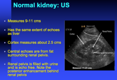
kidney should have same echogenicity as what? central echoes in the kidney are from what? why is the renal pelvis echo free?

answer
can see renal pelvis, cortex, medulla, can measure length of kidney, if there's fluid you will see posterior enhancement (echoes go through fluid as opposed to tissues or fat)
question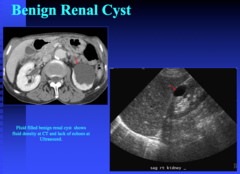
what are the 3 criteria for diagnosing a simple cyst on US? what does a cyst look like on post contrast CT?

answer
on US, *cyst has to be anechoic, pencil thin wall, and through transmission* (the echoes go right through it) that's what classifies a simple cyst but there can be variation to that. may see thick wall or see echoes inside post contrast CT you see hypodensity (fluid)
question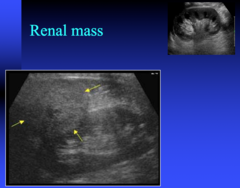
renal mass on US

answer
renal cortex --> you're seeing a large mass (renal cancer)
question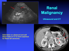
what does RCC look like on US and CT?

answer
on US - arrows point to large mass on CT you see a large heterogenous cancer there's a lot of gray zone in renal masses where cysts can become complex before they're obvious cancers.
question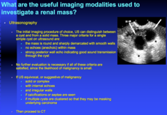
what is the initial imaging procedure of choice to investigate a renal mass? what do you use next for further investigation?

answer
US - can easily see if it's a cyst or not if it's not a cyst then you go onto CT. if US is equivocal or suggestive of malignancy (sold/complex mass irregular walls, calcifications) then you proceed to CT
question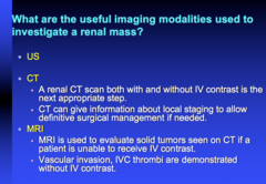
what type of CT should you get to investigate a renal mass? what type of info can this give you? when do you do an MRI?

answer
US *renal CT both with and without contrast. have to do a 2 phase CT*. CT can give info about local staging if surgery management is needed MRI used infrequently. CT is the work horse for majority of renal masses. MRI used if CT has issues (equivocal, certain cystic lesions better evaluated on MRI or vascular invasion)
question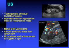
describe how RCC looks like on US

answer
solid mass on US cysts anechoic isoechoic, hypoechoic, hyperechoic are terms used to describe mass on US renal cell cancer - isoechoic mass from upper pole on this example. if it did not deform the pole you may miss an isoechoic mass on US because if it's the same echogenecity as the rest of the cortex you may not see it's a separate mass this is a mass - no posterior wall enhancement and it has same echogenecity as rest of cortex
question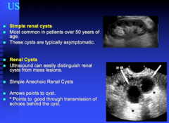
simple renal cysts are common over what age? what else can look like cysts and how do you distinguish the 2?

answer
lymphomas can look dark like cysts but they will not have through transmission - be careful what you call a cyst
question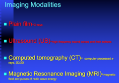
4 imaging modalities for kidney

answer
CT is nice - you can do 2D / 3D imaging
question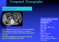
Hounsfield Units

answer
everything on CT is based on Hounsfield Unit - that's how we can distinguish bright bones from soft tissue from fat and from air. those 34 air is -1000, dense bone is +1000 water is 0 cyst = 0-10 is always a cyst can use that to see if it stays that way or if it goes up
question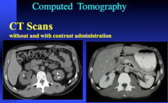
CT with and without contrast - what are the enhancement phases you can use to eval the kidney?

answer
always do CT with and without contrast (with stone you can do non contrast CT). but if there's a question if something is a mass, always do with and without contrast bc you want to see how much something enhances after contrast so you want a pre-contrast Hounsfield Unit and a post-contrast Hounsfield Unit bright things = calcifications post-contrast kidneys are enhancing enhancement has several phases, it's not just post-contrast exam and you're done. early on there's a portal venous phase and then there's a nephrographic veins. if you want to evaluate the kidneys you do a nephrographic phase which is 60-90 sec after contrast injection if a patient comes to ER and you order a CT, it's always done in a portal venous phase bc liver is biggest organ and we eval this first. if something is found in the kidneys, they're often brought back to better eval that because it's a different phase to look at the kidneys
question
kidney is located where & surrounded by what? what part of the kidney enhances with IV contrast? what does this part of the kidney look like in the nephrogenic phase? what can be seen as the contrast is excreted from the kidneys? Renal vein drains where?
answer
renal cortex enhances with IV contrast in nephrographic phase, the contrast has not been excreted and renal pelvis will be dark- that's when you want to eval kidneys
question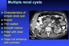
simple renal cyst criteria do they enhance with contrast?

answer
cortical cysts are dark Hounsfield Unit will be 0-10 round thin walled smooth interior filled with clear liquid *does not enhance with contrast* if this was 40 Hounsfield Unit then you want to do a non-contrast to see if it's already 40 before hand (hyper dense cyst) or not. thats when you do further work up
question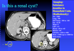
what is the HU for fat? what is the most common fatty lesion in the kidney? what other renal pathology can have microscopic fat?

answer
1st thing - do Hounsfield Unit. turns out to be -40 (fat range) most common fatty lesion in the kidney is angiomyolipoma renal cancers can have microscopic fat
question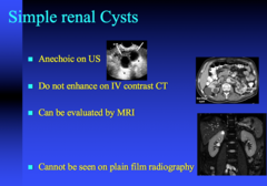
what do simple renal cysts look like on US, CT, MRI, Xray? do they contrast enhance?

answer
through transmission - echoes are going right through it dark hypodense lesions on CT - cysts on MRI T2 weight image the cysts are bright cysts should not enhance on CT cysts are not seen on plain film
question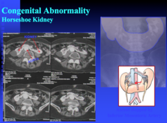
horseshoe kidney on CT horseshoe kidney is prone to what 4 things?

answer
can be seen on CT often present for other reasons - i.e. they want to donate their kidney, trauma horseshoe kidneys are prone to *urine stasis* leading to *stone formation --> infection --> cancer*
question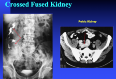
cross fused kidney

answer
on the right side you have 2 kidneys can have pelvic kidney
question
adult polycystic disease - hepatic and renal on CT

answer
hepatic cysts, renal cysts, aneurysm
question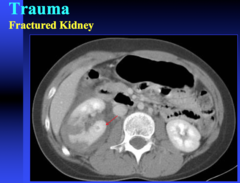
fractured kidney on CT what contrast phase is used to see a fractured kidney on CT?

answer
most often in the ER setting we do a CT in *portal venous phase* for fractured kidney can eval perinephric blood
question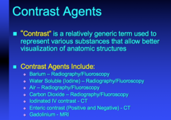
what type of contrast is used for plain films, CT, and MRI?

answer
oral and IV contrast oral = barium for plain films. water soluble contrast. can also use air in some of the studies, CO2 in some interventional studies most of the time we use iodinated IV contrast in CT - can have allergic rxns gadolinium for MRI today there's no US contrast
question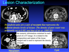
what increase in HU do you need to be worried about a mass?

answer
can characterize that lesion meaning is this a simple cyst, something we need to worry about, do they need surgery etc Hounsfield Units measure the different density levels of tissues. density of pure water is 0 HU left pics: on non contrast scan, it's measuring 22 HU and post contrast it goes up to 64 HU. if it's greater than 10-12 enhancement difference, you need to worry about it. could be a solid mass right pics: 55 HU hyperdense cysts non contrast, post contrast the HU goes to 62. not much enhancement so this is just a hyper dense cyst. HU pre and post is how you characterize a lesion within the kidney
question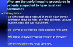
what is the diagnostic procedure of choice for a primary renal tumor? what 5 things can it tell you? what info can MRI tell you?

answer
CT = workhorse US can be a screening cyst to see if something is cystic or not MRI used if a patient has renal dysfunction or allergy to contrast you can use MRI to see if it's a cyst or not IVP and angio not used too much anymore
question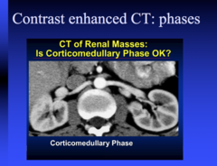
arterial phase, portal venous phase, nephrogenic phase, delayed phase

answer
arterial phase = 25-30 sec after contrast portal venous phase = 60 sec after contrast nephrographic phase = 90 sec after contrast delayed phase = 5 min after contrast injection (eval collecting systems) we don't like to do these phases bc of radiation
question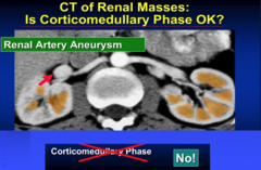
what % of the kidney is enhanced in portal venous phase? what is the best phase to eval kidney?

answer
portal venous phase is 60 sec after contrast injection, looking at liver primarily. in the kidneys you can see cortex enhancing but medulla has not enhanced yet. this is not right phase for evaluating the kidneys bc only 50% of kidneys are being enhanced in this case best phase to eval kidney = nephrographic (after 90 sec of contrast injection) renal artery aneurysm is seen in this phase - can see vascular lesion in this phase but it's not for the kidney
question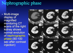
nephrogenic phase is used to eval what?

answer
nephrographic phase - if there's a renal lesion you want that phase
question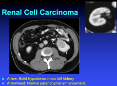
what does RCC looks like on CT?

answer
large mass this is not a cyst bc this is a solid lesion, you would know that if you do HU it's darker than the rest of the cortex but not dark enough for a cyst
question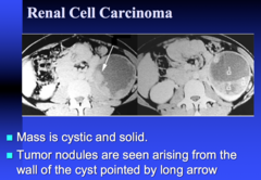
cystic RCC lesion on CT

answer
not just solid lesion, can be cystic as well can be complex cystic which makes it hard to read renal scans this is nodular and enhancing. this is solid and cystic mass that's renal cancer
question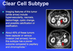
RCC clear cell subtype what does it look like on CT? why is the prognosis worse than chromoprobe or papillary?

answer
different varieties of renal call carcinomas clear cell can cause vascular invasion (left renal vein)
question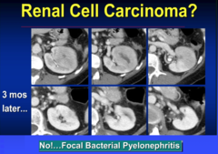
what else can be confused with RCC on CT?

answer
everything that's solid is NOT cancer this looks solid and heterogeneous. this turned out to be infection bc it resolved 3 mo later --> focal bacterial pyelo. can be confused with renal cancer
question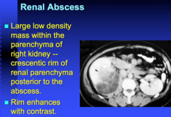
appearance of renal abscess on CT

answer
renal abscesses will be dark and heterogeneous
question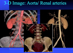
what can be imaged 25-30 seconds after contrast injection? what can be imaged 5 minutes after contrast injection?

answer
vascular eval can be done nicely on CT arterial phase -25-30 seconds after contrast injection. can get images of aorta and eval vessels if you delay injection to 5 minutes you can get collecting system as well you can do both arterial and collecting system in same exam UT urogram = non contrast, nephrographic phase, and delayed phase to look at collecting system
question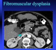
fibromuscular dysplasia is a cause of HTN in what patient population?

answer
often a cause of HTN in middle aged women nicely seen on CT where you see irregular vessels
question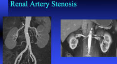
renal artery stenosis

answer
another cause of HTN can do MRI or CT to eval can be treated using a stent - can be done at same time or at a different time
question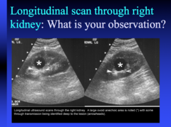
simple cyst on US

answer
simply cyst on US = anechoic (dark), through transmission (echoes go right through it), pencil thin wall
question
most common cause for painful hematuria? what's the best way to image it?

answer
*calculi in GI tract* - best imaging modality for eval is *unenhanced helical CT* high incidence here bc of limestone
question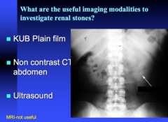
imaging modalities to investigate renal stones when can US be used?

answer
can see psoas and fat outlining the kidney no contrast US? only if you thought they had obstruction (hydronephrosis)
question
non contrast CT has greater sensitivity than what for ureteral stone? non contrast CT has better sensitivity or specificity? what are 4 pros of using non contrast CT? what stones can't be picked up by contrast CT?
answer
CT - can see hydronephrosis, how big the calculi are
question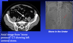
what is a feature of hydronephrosis on CT? what's stone protocol

answer
*hydronephrosis - left kidney has fat stranding*, it gets enlarged and there's fluid in the pelvis if you follow the ureter down you can see where the stone is -normal CT scan slice = 5 mm -stone protocol = 3 mm
question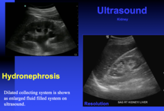
what are the limitations of using US to see obstruction causing hydronephrosis?

answer
US - can see hydro easily they may not have hydronephrosis but they may have a stone that's non obstructive but still in the mid ureter. *on US you can only eval the kidney and proximal renal pelvis, you can't go below this bc of fat* US best just for the kidney level but CT you can go all the way down and see it
question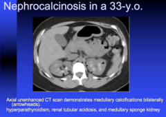
what are 3 cases of nephrocalcinosis? what's the most common?

answer
these don't look like renal stones this is more for congenital reasons medullary calcifications ddx - most often from medullary sponge kidney, can also get from hyperpara and RTA
question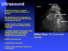
US is preferred to CT in what 2 patient groups? what types of stones does it detect? appearance of stone on US what types of stones does it have difficulty detecting?

answer
on US - stones will be bright and will keep the echo from going. so you'll see a shadow behind it. anything that obstructs the movement of sound will cause a shadow. typically example of renal stone on US cysts will let echoes go through while either fat/stone will not let the echo go through and cause a shadow
question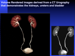
CT urogram what drug can you give to force contrast to go up into the collecting system and fill up the ureters? CT urogram is usually for what kidney pathology?

answer
CT urogram, 5 min after contrast injection also give lasix to force contrast to go into collecting system and fill up ureters bilaterally this is not done for stone, more for transitional cell carcinoma or lesions in the ureters
question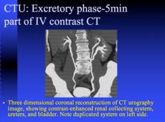
CT urogram can be used to detect which congenital anomalies?

answer
congenital anomalies - duplicated collecting system, ectopic insertion of ureter which may cause infection
question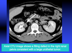
axial CTU is most often done for what kidney pathology?

answer
*transitional cell cancer is the most common reason this is done* soft tissue mass causing a filling defect
question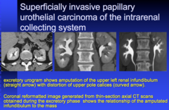
what's an excretory urogram?

answer
can see defects on contrast exam this is called excretory urogram, delayed post 5 min
question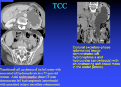
TCC on imaging

answer
this lesion caused upstream hydro in the left kidney, TCC
question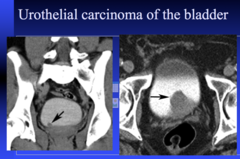
what's another name for TCC?

answer
urothelial cancer and TCC are used synonymously
question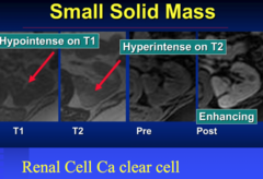
what are 3 reasons to use MRI? what type of cancer can it be used to view and why?

answer
MRI useful when 1. CT equivocal 2. lesion is getting larger 3. some subtypes of RCCs clear cell is easy = heterogenous, fairly enhancing mass *subtype called papillary renal cell may not enhance as much on CT (may only have 12-15 HU change)* there are some subtypes of papillary that are more aggressive than others. then you go to MRI to further eval these lesions MRI used more where it's gray zone on CT
question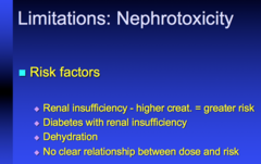
what are 3 risk factors for nephrotoxicity due to contrast? what's the relationship between dose and risk?

answer
nephrotoxicity is a problem bc of contrast in both CT and MRI can't give contrast to everyone half of the dose doesn't matter
question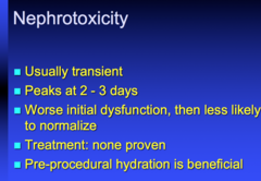
describe the nephrotoxicity that occurs with contrast what's the treatment? how do you prevent it?

answer
typical nephrotoxicity that occurs with CT contrast is usually not a big problem
question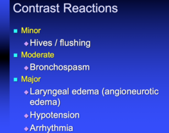
what are minor (1), moderate (1), and major (3) contrast reactions?

answer
minor = hives/flushing can get bronchospasm most important = arrhythmia 2nd time they're exposed to contrast it can be major. never give them contrast again if they had a rxn the first time
question
how are renal masses usually detected? what intervention do you do when you find simple cyst? incidental RCCs have lower stage of what?

answer
general renal masses - most incidental on US, CT, MRI mosts are simple cysts and require no action RCCs being caught at very low stage - have to eval and follow
question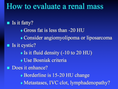
how to eval a renal mass?

answer
always check HU - fatty or not, fluid, etc Bosniak criteria - how you grade cystic lesions on CT or US. higher Bosniak = more likely to be cancer. 3 and higher they would get surgery. papillary RCC not simple mets, IVC clot, lymphadenopathy will enhance
question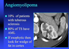
angiomyolipoma is associated with what? if exophytic what do you look for?

answer
angiomyolipoma - 10% of patients with tuberous sclerosis 80% of TS will have angiomyolipoma


