Skeletal Radiology IV: Benign Bone Tumors and Tumor Like-Conditions Part 1 – Flashcards
Unlock all answers in this set
Unlock answersquestion
What is a bone island?
answer
A circumscribed focus of cortical bone wtihin cancellous bone. It is usually small and under *1cm* in diameter. Can have a brush border. Is very common. Not symptom producing.
question
You find what appears to be a bone island. What else could it be?
answer
*Blastic Metastasis* Also, prostate cancer metastatic to bone, sesamoid bones, bowel contents (like swallowed pills)
question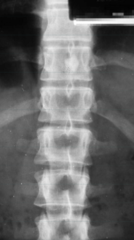

answer
Bone Island
question

answer
Bone Island
question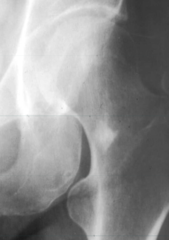

answer
Bone Island (Note the brush border)
question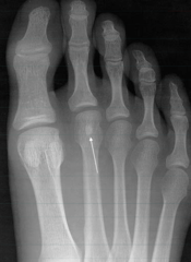

answer
Either an accessory sesamoid or a bone island on the 2nd toe. It really doesn't matter which. The other 2 on the first toe are both sesamoid bones.
question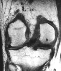

answer
Bone Island (On MRI - Note it looks black)
question
What is Osteopoikilosis?
answer
Spotted bone disease. It is an autosomal dominant condition characterized by hundreds of bone islands which tend to cluster around large joints.
question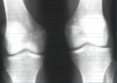

answer
Osteopoikilosis
question
Fill this out for Osteopoikilosis. *Number of Lesions*: *Size and Uniformity of Lesions*: *Symptoms*: *MRI Findings*: *Lab Findings*: *Change over Time*:
answer
*Number of Lesions*: Hundreds *Size and Uniformity of Lesions*: Uniformly small (1cm or less) *Symptoms*: None *MRI Findings*: MRI has no signal *Lab Findings*: None *Change over Time*: No change with serial films!!!!
question
Fill this out for Blastic Mets. *Number of Lesions*: *Size and Uniformity of Lesions*: *Symptoms*: *MRI Findings*: *Lab Findings*: *Change over Time*:
answer
*Number of Lesions*: Usually multiple *Size and Uniformity of Lesions*: Varying sizes *Symptoms*: Bone pain, increasing in severeity *MRI Findings*: MRI findings with it being very high on T2 *Lab Findings*: Present (High PSA) *Change over Time*: Growth, additional lesions over time
question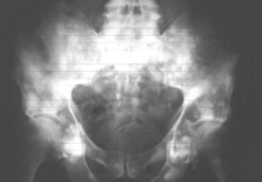

answer
Osteoblastic Metatasis
question
We see Osteopoikilosis. Before we make out diagnosis we must rule out what condition?
answer
Osteoblastic Metastasis
question
What is a Hamartoma?
answer
A *benign* growth made up of an abnormal mixture of cells and tissues normally found in the area of the body where the growth occurs.
question
What are osteoma?
answer
Comprised of normal bone constituents that grow only on skull or sinuses (because they undergo intramembranous bone formation as opposed to endochondral)
question
Osteomas are insignificant except for when....
answer
They may be palpable, can block a sinus outflow, or Gardner's Syndrome.
question
What are the symptoms of Gardner's syndrome
answer
*1.* Osteomas (Palpable) *2.* Colon Polyps (Which can undergo metastatic change) *3.* Skin Lesions
question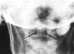

answer
Osteoma
question

answer
Osteoma
question

answer
Osteoma
question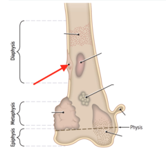

answer
Osteoid Osteoma
question
What is an osteoid Osteoma?
answer
A small vascular tumor which is *benign*. However, tumor is quite painful (especially at night). Can affect any bone including the spine. When affecting spine, it loves the posterior elements. *Dramatically relieved by NSAIDS*
question
What is the prognosis of Osteoid Osteomas?
answer
Self limiting. May go away on its own.
question
What age range is affected by Osteoid Osteoma?
answer
Age 10-25
question
What do Osteoid Osteomas look like radiographically?
answer
Small radiolucency (called a *nidus*) which can have central calcification. Nidus can be intracortical or intramedullary. Solid reactive new bone may be more noticeable than the tumor itself.
question
Is a lucent nidus or a calcified nidus more associated with an older tumor?
answer
Calcified
question

answer
Osteoid Osteoma
question

answer
Osteoid Osteoma
question

answer
Osteoid Osteoma
question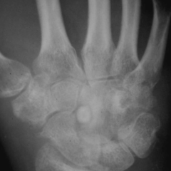

answer
Osteoid Osteoma (Notice it is surrounded by the bodys reaction to it)
question
How do we treat Osteoid Osteoma
answer
*1.* Do nothing... may go away on their own. *2.* If painful, use percutaneous radiofrequency Ablation. Under CT guidance, a needle is inserted into nidus for biopsy. Nidus replaced with probe. Tumor is heated to kill the tumor cells.
question
Osteoid Osteomas tend to occur in the (anterior/posterior) segment of the spine.
answer
Posterior
question
Osteoid Osteomas take approximately how long to go away on their own?
answer
2 years
question
What is the differential diagnosis for an osteoid osteoma?
answer
*1.* Healing Stress fracture (will also have pain and reactive sclerosis. No nidus and might see a fracture line) *2.* Brodie's Abscess (will also have pain, a small lucency, but no reactive sclerosis)
question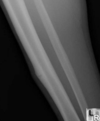

answer
Healing Stress Fracture
question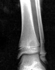

answer
Brodie's Abscess
question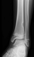

answer
Fibrous Cortical Defect
question
Painful tiny benign tumors with big reactive sclerosis are called....
answer
Osteoid Osteomas
question
A giant osteoid osteoma is called...
answer
Osteoblastoma
question
What is an Osteoblastoma?
answer
Histologically related to osteoid osteoma. These are larger and expansile. Pain is less intense but is *not* worse at night and *not* dramatically relieved by nsaids.
question
What age group is most affected as osteoid osteomas
answer
10-25
question
Are osteoblastomas or osteoid osteomas more likely to affect the spine?
answer
Osteoblastomas. 40% of them affect the spine (most in posterior elements)
question
Neurological complications are more common in (osteoblastomas/osteoid osteomas).
answer
Osteoblastomas (because they are more expansile)
question
What percentage of Osteoblastomas affect the spine?
answer
40%
question
Fill this out for Osteoid Osteomas *Age Affected*: *Size of Lesion*: *Expansile*: *Soap Bubbly*: *Bones Affected*:
answer
*Age Affected*: 10-25 *Size of Lesion*: small (less than 1cm) *Expansile*: No *Soap Bubbly*: No *Bones Affected*: Any
question
Fill this out for Osteoblastomas *Age Affected*: *Size of Lesion*: *Expansile*: *Soap Bubbly*: *Bones Affected*:
answer
*Age Affected*: 10-25 *Size of Lesion*: Larger (larger than 2cm) *Expansile*: Yes *Soap Bubbly*: Yes *Bones Affected*: Any, but 40% are in the spine (mostly posterior elements)
question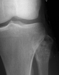

answer
Osteoblastoma (Appears aggressive)
question
In the spinal canal, where is the cord? What is the significance?
answer
It is more anterior. So tumors which fill in posterior spaces tend to not cause neurological conditions.
question
What is the most common benign bone tumor?
answer
Osteochondroma
question
What are osteochondromas?
answer
Benign growths at the corner of the growth plates. They are encapsulated by a cartilage cap. Osteochondromas have both a bone and cartilage component.
question
Osteochondromal lesions (farther from/closer to) the axial skeleton are more likely to become malignant.
answer
Closer to
question
When do Osteochondromas develop?
answer
Develop early in childhood and are caused by trauma to epiphyseal plate.
question
Osteochondromas grow until when?
answer
Until the child stops growing.
question
Osteochondromas do/do not affect the spine.
answer
*Do*
question
Osteochondromas tend to point (towards/away) from the closest joint.
answer
Away from
question

answer
Osteochondroma?
question
Osteochondromas are typically (symptomatic/asymptomatic)
answer
Asymptomatic
question
What are the two types of osteochondromas?
answer
Sessile (flat base) and pedunculated (stalked)
question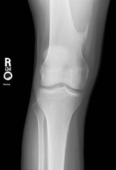

answer
Pedunculated Osteochondroma
question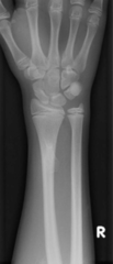

answer
Sessile Osteochondroma
question
What arethe "nicknames" for the two types of Osteochondromas?
answer
*Sessile*: Cauliflower Exostosis *Pedunculated*: Coat Hanger Exostosis
question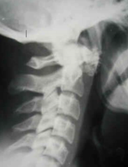

answer
Osteochondroma of the C1. Sorry. This is a cheapshot. There is no way for you to determine what this could be. Could be ligament or muscle ossification. Just be aware it could be this.
question
Osteochondromas are asymptomatic unless...
answer
Cosmetic/palpable, mechanical irritation, or fracture *1% of them turn into malignant change*. Its the same as a mole on your skin. Could become cancerous sure. But unlikely.
question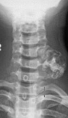

answer
Osteochondroma Sessile
question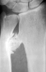

answer
Pedunculated Osteochondroma causing mechanical irritation. It formed a pseudo-joint (adventitious joint) which underwent degenerative joint disease. Note presence of osteophytes.
question
How many osteochondromas become malignant?
answer
Less than 1%. Like the chances of a mole.
question
When do we worry about osteochondromas becoming malignant? (symptoms)
answer
Osteochondroma continues growing after he has stopped growing and has developed pain.
question
What is one way to tell the malignant potential of Osteochondromas?
answer
MRI assessing the cartilage cap. Cartilage is the main offender in this condition. Cartilage cap more than ; 2cm thick is considered for malignancy.
question
MRI hallmark of malignant degeneration for osteochondromas is thickening of cartilage cap. What thickness indicates malignant change?
answer
;2cm
question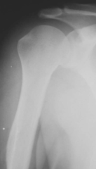

answer
Normal. That mass on the humerus is the deltoid process.
question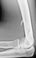

answer
Supracondylar process/spur. Not to be confused with osteochondromas. We know this because the spur is facing towards the joint.
question
What is another word for osteochondroma?
answer
Exostosis
question
What is HME?
answer
Hereditary Multiple Exostosis (or Hereditary Multiple Osteochondroma) Autosomal dominant condition characterized by dozens of exostosis lesions.
question
Regarding Fibroxanthomas, what is the name for one that is small and one that is large.
answer
*Small*: Fibrous Cortical Defect *Large*: Non-Ossifying fibroma
question
Compare and contrast Fibrous cortical defects and nonossifying fibromas. *Size*: *Soap Bubbly?*: *Path Fx?*: *Clin Significance*: *Spinal?*:
answer
*Size*: FCD is 3cm *Soap Bubbly?*: No to FCD and yes to NOF *Path Fx?*: No to FCD and Yes to NOF *Clin Significance*: No big deal for FCD. Path Fx possible in NOF *Spinal?*: No for both.
question
Where are you more likely to see monostotic fibrous dysplasia?
answer
Proximal Femur and Rib
question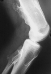

answer
HME/HMO Hereditary Multiple Exostosis
question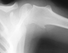

answer
HME/HMO
question

answer
HME/HMO
question
What is the chance of malignant change with HME?
answer
5-25%
question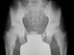

answer
HME/HMO. Notice the only sign is widened femoral necks.
question
In osteochondroma, malignant transformation occurs in what portion of? What will it turn into?
answer
Cartilage. Chondrosarcoma.
question
What is the MC benign bone tumor of the small, tubular bones of the hands and feet?
answer
Enchondroma
question
Enchondroma is the MC bengin bone tumor of...
answer
Small tubular bones of hands and feet
question
Tell me about Enchondromas.
answer
Potentially expansile with a cartilage matrix which can calcify. *Does not affect spine and usually symptomatic*.
question
Under what conditions are enchondromas potentially symptomatic?
answer
Path fracture or malignant transformation
question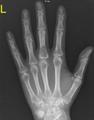

answer
Enchondroma
question

answer
Enchondroma
question

answer
Enchondroma (Popcorn calcification)
question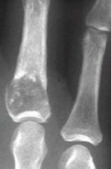

answer
Enchondroma (Popcorn calficiation and path fracture)
question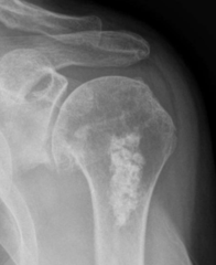

answer
Enchondroma (Notice popcorn calcification) This patient has an osteophyte on the inferior of the humeral head. Probably why they came into get an xray.
question
Any time you see an enchondroma, what two things must you consider for your differential diagnosis?
answer
*1.* Medullary Bone Infarct *2.* Low grade chondrosarcoma
question
Multiple Enchondroma is called...
answer
Ollier Disease
question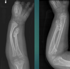

answer
Ollier Disease
question
How do you get Ollier disease?
answer
You are born with it but it is *not hereditary*.
question
What is a quick way to assess a child's age if you have an x-ray of their hand?
answer
children *generally* gain one new ossified carpal bone per year. This should tell you imprecisely what their bone age should be. Can help you assess development.
question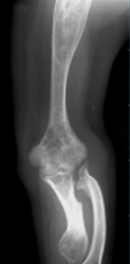

answer
Ollier Disease. Note the many enchondromas. This disease has caused deformity in the proximal limb.
question
What is Maffucci Syndrome?
answer
Ollier disease combined with soft tissue hemangiomas. Soft tissue is usually difficult to see on x-ray but these hemangiomas are calcified into phleboliths. In addition to endochondromas, phleboliths are pathognomonic for Maffucci syndrome. (Usually if phleboliths are anywhere other than pelvis)
question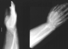

answer
Maffucci Syndrome
question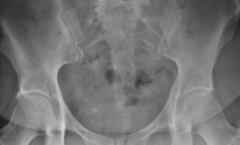
What is this?

answer
Phleboliths. Small calcifications in the veins.
question
What is the most common benign bone tumor of the hands and feet?
answer
Enchondromas
question
What is the cancer risk with patients who have enchondromas?
answer
*Rare* Enchondromas have a 1-5% chance. However, Ollier is 25% and Maffucci is 50%. Don't get too focused on memorizing.
question
There is a higher chance for enchondroma lesions the closer you get to....
answer
Axial skeleton.
question
What is the ddx for enchondromas?
answer
*1.* Medullary infarct *2.* Low grade chondrosarcoma.
question
What are Chondroblastomas?
answer
Rare cartilage tumors affecting children and young adults. (Mean age is 20) Arises in the epiphysis. *Only tumor to do this*. Usually affects lower extremity.
question
The only tumor to arise in the epiphysis is the...
answer
Chondroblastoma.
question
Chondroblastomas typically affect what?
answer
Lower extremities. Half are around the knees.
question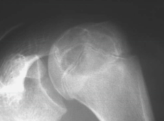

answer
Chondroblastoma
question

answer
Chondroblastoma
question
What are the symptoms of Chondroblastomas?
answer
Joint pain
question
What is the only true epiphyseal tumor
answer
Chondroblastoma. Only one that starts there.
question
T/F: Cartilage tumors do not like the spine?
answer
True.
question
Clinically, patients with HME presence how physically?
answer
Short stature and visible palpable limb deformities.
question
T/F: Fibrous Cortical Defects can sometimes have pain.
answer
False. No pain. This is unlike osteoid osteomas.
question
Majority of Fibrous Cortical Defect is seen in.....
answer
Metaphyseal *cortex* of long bones
question
Patient has no pain.

answer
Fibrous Cortical Defect
question
Fibrous Cortical Defects are under ___ cm.
answer
3 cm
question

answer
Fibrous Cortical Defect
question
T/F: Fibrous Cortical defects never affect the spine.
answer
False. They typically do not affect the spine.
question
How many cm's (generally) are 1 inch?
answer
2.5 (2.54 specifically) is 1 inch. You need to know this for patients. "Your tumor is 2.5cm in length" tells little to most patients.
question
Non-Ossifying Fibroma are most clinically significant due to...
answer
Pathological fractures.
question
How large are non-ossifying fibroma?
answer
Over 3cm in length.
question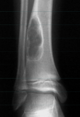

answer
Non-ossifying Fibroma
question
Tell me about the physical appearance of non-ossifying fibroma.
answer
Soap bubbly and surrounded by a sclerotic rim. It is slightly expansile and is over 3cm in length.
question

answer
Non-Ossifying Fibroma
question
Is ESR accelerated in heart failure?
answer
No
question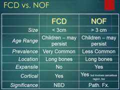
Compare and contrast the size, age range, prevalence, location, expansile nature, cortical involvement, and clinical significance of FCD vs NOF and then look at the picture to see if you got it right.

answer
Yay
question
Where do we often find Non-ossifying Fibromas?
answer
Metaphysis of long bones
question
Non-Ossifying Fibromas are generally (painful/not painful)
answer
Not painful
question
T/F: Fibrous Dysplasia MUST develop in childhood. If it doesn't, you will never get it.
answer
True
question
What are the 4 forms of Fibrous Dysplasia?
answer
*1.* Monostotic - most common *2.* Polyostotic (With endocrinopathy you get diabetes myellitus, cushing syndrome, and precocious puberty-McCune-Albright) *3.* Craniofascial *4.* Cherubism
question
Tell me about Monostotic Fibrous Dysplasia
answer
Very common, painless, affects ribs mostly (but also long bones), and develops during childhood.
question
What do monostotic Fibrous Dysplasia look like radiographically?
answer
Lesions range from lucent to opaque or in-between. (Ground glass or frosted mug appearance). Often have thick rim of sclerosis called a "rind" sign. Can be expansile.
question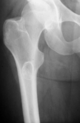

answer
Rind sign, ground glass, and location tell us this is likely *Fibrous Dysplasia*
question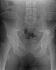

answer
Great example of Rind sign. Fibrous Dysplasia.
question
What classification system can we use to assess fracture risk?
answer
Mirel
question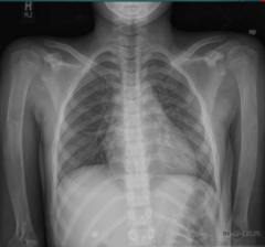
Ignore humerus for now.

answer
Right 2nd rib and posterior portion of left 4th rib shows a monostotic Fibrous Dysplasia that is expansile.
question
Tell me about Polyostotic Fibrous Dysplasia
answer
Many tumors of fibrous dysplasia. Can happen with or without endocrinopathy. Asymmetrical and 50% of patients have cafe-au-lait spots that look like the Coast of Maine (Small islands).
question
What do the cafe-au-lait spots look like in Polyostotic Fibrous Dysplasia?
answer
"Coast of maine". Large "landmass" with many "islands"
question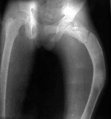

answer
Shepherd's crook deformity
question
What is a Shepherd's crook deformity?
answer
Proximal femur deformity due to fibrous dysplasia. Usually coxa vara and less than 120. (normal femoral neck angle is 120-130)
question
Normal femoral neck angle is how many degrees?
answer
120-130
question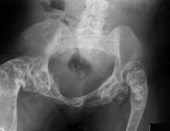

answer
Shephard's Crook
question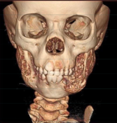

answer
Cherubism


