Derm Pics (Fitzpatrick’s 6/7th ed & Bates) – Flashcards

Unlock all answers in this set
Unlock answersquestion")
Impetigo (BViral PP5)
answer
Identify
question")
Impetigo (BViral PP6)
answer
Identify
question
Impetigo (BViral PP6)
answer
Identify
question (BViral PP7)")
Streptococcal Pyoderma (ecthyma) (BViral PP7)
answer
Identify
question")
Impetigo (BViral PP8)
answer
Identify
question")
Impetigo (BViral PP 9)
answer
Identify
question")
Bullous Impetigo (BViral PP11)
answer
Identify
question")
Bullous Impetigo (BViral PP12)
answer
Identify
question")
Bullous Impetigo (BViral PP12a)
answer
Identify
question")
Bullous Impetigo blistering dactylitis (BViral PP13)
answer
Identify
question")
Scalded Skin Syndrome (BViral PP15)
answer
Identify
question")
Scalded Skin Syndrome (BViral PP16)
answer
Identify
question
Scalded Skin Syndrome (BViral PP16)
answer
Identify
question")
Folliculitis (BViral PP18)
answer
Identify
question
Folliculitis (BViral PP18)
answer
Identify
question")
Furuncle (BViral PP20)
answer
Identify
question")
Furuncle (BViral PP21)
answer
Identify
question
Furuncle (BViral PP21)
answer
Identify
question")
Abscess (BViral PP22)
answer
Identify
question")
Multiple furuncles (BViral PP23)
answer
Identify
question")
Carbuncle (BViral PP24)
answer
Identify
question")
Cellulitis (BViral PP28)
answer
Identify
question
Cellulitis Traditionally, simple cellulitis not requiring hospital admission has most commonly been treated with a penicillinase-resistant penicillin (e.g., dicloxacillin), or an oral cephalosporin (e.g., cephalexin) (see Box 178-2). Cephalexin Dicloxacillin (Fitzpatricks 8e Box 178-2; BViral PP29)
answer
How is this out patient condition treated
question")
Cellulitis (BViral PP29)
answer
Identify
question
Cellulitis Legend: Surgical excision wound infection: MSSA Surgical wound became painful and tender 7 days after excision of squamous cell carcinoma; soft tissue (cellulitis) is seen adjacent to the wound margin. Necrotic tissue is seen in the base (BViral PP30)
answer
Identify
question
Acute lymphangitis of forearm - Cellulitis Acute lymphangitis of forearm: S. aureus A small area of the cellulitis on the volar wrist with a tender linear streak extending proximally up the arm; the infection spreads from the portal of entry within the superficial lymphatic vessels (BViral PP31)
answer
Identify
question")
Cellulitis 1st drug of choice: Ampicillin/sulbactam Ticarcillin/clavulanate Piperacillin/tazobactam Imipenem/cilastatin, meropenem Alternative: Vancomycin Clindamycin Linezolid (Fitzpatricks 8e Box 178-2; BViral PP32)
answer
How are hospitalized patients with this condition treated?
question")
Cellulitis Refractory, high likelihood of MRSA infection: Vancomycin Linezolid (Fitzpatricks 8e Box 178-2; BViral PP32)
answer
How are patients with this condition and with a high likelihood of MRSA treated?
question")
Cellulitis (BViral PP32)
answer
Identify
question")
Erysipelas Penicillin Va Intramuscular procaine penicillin Amoxicillin Vancomycin (Fitzpatricks 8e Box 178-2; BViral PP35)
answer
Treatment for this condition
question
Erysipelas Penicillin Va Intramuscular procaine penicillin Amoxicillin Vancomycin (Fitzpatricks 8e Box 178-2; BViral PP35)
answer
How is this condition treated?
question")
Infectious Intertrigo (BViral PP35)
answer
Identify
question
Infectious Intertrigo (BViral PP35)
answer
Identify
question
Infectious Intertrigo Legend: Intergluteal intertrigo: group A streptococcus A painful moist erythematous plaque in a male with intertriginous psoriasis, with foul odor. Infection resolved with penicillin VK. (BViral Lecture PP38)
answer
Identify
question")
Erythrasma (BViral Lecture PP40)
answer
Identify
question
Necrotizing Fasciitis Short of open exploration, no test is specific for necrotizing infection, but several laboratory, microbiologic, histopathologic, and imaging studies have been shown to be useful in differentiating bacterial necrotizing SSTI from non-necrotizing infections or noninfectious causes of soft-tissue necrosis (see Box 179-1) (Fitzpatricks 8e CH 179; BViral Lecture PP41)
answer
How is this condition diagnosed
question")
Atopic Dermatitis (Marlin D&E Lecture PP20)
answer
What condition has this distribution pattern?
question
Atopic Dermatitis is treated with: Minimize skin irritation Soaps, clothing Lukewarm baths Corticosteroids-topical/oral Control pruritis-antihistamines Skin lubrication Treat 2dary bacterial infection Avoidance of food allergens Referral to Allergist (Marlin D&E Lecture PP22;28)
answer
What is the treatment for this condition?
question
Atopic Dermatitis is treated with: Minimize skin irritation Soaps, clothing Lukewarm baths Corticosteroids-topical/oral Control pruritis-antihistamines Skin lubrication Treat 2dary bacterial infection Avoidance of food allergens Referral to Allergist (Marlin D&E Lecture PP23;28)
answer
What is the treatment for this condition?
question
Legend: Childhood atopic dermatitis A typical localization of atopic dermatitis in children is the region around the mouth. **In this child, there is lichenification and fissuring and crusting. (Marlin D&E Lecture PP23)
answer
What unique feature** is identifiable in this condition?
question")
Infant Atopic Dermatitis (Marlin D&E Lecture PP 24)
answer
Identify
question
Atopic Dermatitis is treated with: Minimize skin irritation Soaps, clothing Lukewarm baths Corticosteroids-topical/oral Control pruritis-antihistamines Skin lubrication Treat 2dary bacterial infection Avoidance of food allergens Referral to Allergist (Marlin D&E Lecture PP25;28)
answer
What is the treatment for this condition?
question
Atopic Dermatitis is treated with: Minimize skin irritation Soaps, clothing Lukewarm baths Corticosteroids-topical/oral Control pruritis-antihistamines Skin lubrication Treat 2dary bacterial infection Avoidance of food allergens Referral to Allergist (Marlin D&E Lecture PP26;28)
answer
What is the treatment for this condition?
question
Atopic Dermatitis is treated with: Minimize skin irritation Soaps, clothing Lukewarm baths Corticosteroids-topical/oral Control pruritis-antihistamines Skin lubrication Treat 2dary bacterial infection Avoidance of food allergens Referral to Allergist (Marlin D&E Lecture PP27;28)
answer
What is the treatment for this condition?
question Topical high potency steroids Antibiotic Tx for infection Systemic steriods Intralesional Triamcinolone (Marlin D&E Lecture PP49;53)")
Dyshidrosis is treated with: Burrows solution (Domeboro) Topical high potency steroids Antibiotic Tx for infection Systemic steriods Intralesional Triamcinolone (Marlin D&E Lecture PP49;53)
answer
What is the treatment for this condition?
question
Dyshidrosis is treated with: Burrows solution (Domeboro) Topical high potency steroids Antibiotic Tx for infection Systemic steriods Intralesional Triamcinolone (Marlin D&E Lecture PP50;53)
answer
What is the treatment for this condition?
question
Dyshidrosis is treated with: Burrows solution (Domeboro) Topical high potency steroids Antibiotic Tx for infection Systemic steriods Intralesional Triamcinolone (Marlin D&E Lecture PP50;53)
answer
What is the treatment for this condition?
question
Dyshidrosis is treated with: Burrows solution (Domeboro) Topical high potency steroids Antibiotic Tx for infection Systemic steriods Intralesional Triamcinolone (Marlin D&E Lecture PP51;53)
answer
What is the treatment for this condition?
question
Dyshidrosis is treated with: Burrows solution (Domeboro) Topical high potency steroids Antibiotic Tx for infection Systemic steriods Intralesional Triamcinolone (Marlin D&E Lecture PP52;53)
answer
What is the treatment for this condition?
question")
Lichen Simplex Chronicus Treatment: Interrupt the Scratch Itch cycle Topical steroids Intralesional Triamcinolone Antihistamines- particularly at night (Marlin D&E Lecture PP56;57)
answer
What is the treatment for this condition?
question")
Lichen Simplex Chronicus Treatment: Interrupt the Scratch Itch cycle Topical steroids Intralesional Triamcinolone Antihistamines- particularly at night (Marlin D&E Lecture PP56;58)
answer
What is the treatment for this condition?
question")
Lichen Simplex Chronicus Treatment: Interrupt the Scratch Itch cycle Topical steroids Intralesional Triamcinolone Antihistamines- particularly at night (Marlin D&E Lecture PP56;59)
answer
What is the treatment for this condition?
question
Atopic Dermatitis: Minimize skin irritation Soaps, clothing Lukewarm baths Corticosteroids-topical/oral Control pruritis-antihistamines Skin lubrication Treat 2dary bacterial infection Avoidance of food allergens Referral to Allergist (Marlin D&E Lecture PP28; Bates pg 197)
answer
What is the treatment for this condition?
question
Infected Atopic Dermatitis: Minimize skin irritation Soaps, clothing Lukewarm baths Corticosteroids-topical/oral Control pruritis-antihistamines Skin lubrication Treat 2dary bacterial infection Avoidance of food allergens Referral to Allergist (Marlin D&E Lecture PP28; Bates pg 197)
answer
What is the treatment for this condition?
question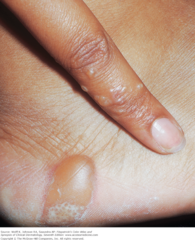
Autosensitization "id Reaction" Treatment with corticosteroids (Marlin D&E Lecture PP54;55)
answer
What is the treatment for this condition?
question
Autosensitization "id Reaction" Caused by release of cytokines as a result of sensitization a primary site Lesions persist until primary problem resolved (Remember treatment: corticosteroids) (Marlin D&E Lecture PP54;55)
answer
Cause of condition? How is it resolved?
question")
Lichen Simplex Chronicus Development of epidermal hyperplasia Secondary to physical trauma Skin becomes highly sensitive to touch (Marlin D&E Lecture PP56;57)
answer
What can this condition be secondary to and what cell condition is developed? Is the skin sensitive? If so to what? If not why?
question")
Lichen Simplex Chronicus Development of epidermal hyperplasia; Secondary to physical trauma Skin becomes highly sensitive to touch (Marlin D&E Lecture PP56;57)
answer
What can this condition be secondary to and what cell condition is developed? Is the skin sensitive? If so, to what? If not why?
question
Autosensitization "id Reaction" Corticosteroids Legend: Prednisone was given for 2 weeks; pruritus and vesiculation resolved.
answer
What can be Rx'd and for what duration?
question
Lesions maculopapular or papulovesicular. Legend: Autosensitization dermatitis ("id" reaction): Bullous (inflammatory) tinea pedis was present and was associated with dermatophytid reaction. Prednisone was given for 2 weeks; pruritus and vesiculation resolved. (Marlin D&E Lecture PP54;55)
answer
What is another name for this condition?
question
Autosensitization "id Reaction": Legend: Bullous (inflammatory) tinea pedis (Marlin D&E Lecture PP54;55)
answer
Describe the larger vesicle
question")
Irritant Dermatitis Prevention of Irritant Dermatitis Removal of the irritant If exposed, wash with water or a neutralizing solution (Marlin D&E Lecture PP5;16)
answer
Identify
question
Irritant Contact Dermatitis Tx: topical or IM corticosteroids Prednisone-tapering dose Antihistamines Wet dressings-Domeboro Soothing baths- Aveeno Watch for 2ary bacterial ifxn (Marlin D&E Lecture PP6;16)
answer
What is the treatment for this condition?
question
Irritant Contact Dermatitis Tx: topical or IM corticosteroids Prednisone-tapering dose Antihistamines Wet dressings-Domeboro Soothing baths- Aveeno Watch for 2ary bacterial ifxn (Marlin D&E Lecture PP7;16)
answer
What is the treatment for this condition?
question
Acute allergic contact dermatitis Legend: Note bright erythema, microvesiculation. At close inspection, a papular component can be discerned. At this stage, there is still sharp margination. (Marlin D&E Lecture PP10)
answer
How can this condition be described?
question")
Acute allergic contact dermatitis (Marlin D&E Lecture PP10)
answer
Identify
question")
Legend: Acute allergic contact dermatitis on the lips due to lipstick (Marlin D&E Lecture PP10)
answer
The patient was hypersensitive to eosin so at this stage what is this condition called?
question")
Allergic phytodermatitis of leg (Marlin D&E Lecture PP12)
answer
Identify
question")
Allergic Contact Dermatitis (Marlin D&E Lecture PP13)
answer
Identify
question")
Allergic Contact Dermatitis Prevention of Irritant Dermatitis Removal of the irritant If exposed, wash with water or a neutralizing solution (Marlin D&E Lecture PP13;16)
answer
How is this condition treated?
question
Allergic phytodermatitis of leg Legend: Linear vesicular lesions with erythema and edema on the calf at sites of direct contact of the skin 5 days after exposure with the poison ivy leaf. (Marlin D&E Lecture PP12;16)
answer
How can this condition be described?
question")
Allergic phytodermatitis of leg Prevention of Irritant Dermatitis Removal of the irritant If exposed, wash with water or a neutralizing solution (Marlin D&E Lecture PP12;16)
answer
How is this condition treated?
question
Allergic Contact Dermatitis Prevention of Irritant Dermatitis Removal of the irritant If exposed, wash with water or a neutralizing solution (Marlin D&E Lecture PP14;16)
answer
How is this condition treated?
question")
Allergic Contact Dermatitis (Marlin D&E Lecture PP14;16)
answer
Identify
question
Allergic Contact Dermatitis Prevention of Irritant Dermatitis Removal of the irritant If exposed, wash with water or a neutralizing solution (Marlin D&E Lecture PP15;16)
answer
How is this condition treated?
question
Allergic Contact Dermatitis (Marlin D&E Lecture PP15;16)
answer
Identify
question
Allergic Contact Dermatitis (Marlin D&E Lecture PP15;16)
answer
Identify
question
Allergic Contact Dermatitis Prevention of Irritant Dermatitis Removal of the irritant If exposed, wash with water or a neutralizing solution (Marlin D&E Lecture PP15;16)
answer
How is this condition treated?
question")
Seborrheic Dermatitis (Marlin D&E Lecture PP31;34)
answer
Identify
question Topical corticosteroids Oral antibiotics if infection (Staph) Removal of crusts in infants using warm oil then shampoo with sulfur shampoo (Marlin D&E Lecture PP31;34)")
Seborrheic Dermatitis Tx: Acute & Maintenance OTC dandruff shampoo x 5-10 mins.-Head & Shoulders or Selsun Cold Tar shampoos-T-Gel, Zetar Ketoconazole cream 2% or shampoo(Nizoral) Topical corticosteroids Oral antibiotics if infection (Staph) Removal of crusts in infants using warm oil then shampoo with sulfur shampoo (Marlin D&E Lecture PP31;34)
answer
How is this condition treated?
question (Marlin D&E Lecture PP31;34)")
Seborrheic Dermatitis Tx: Topical corticosteroids Oral antibiotics if infection (Staph) (Marlin D&E Lecture PP31;34)
answer
How is this condition treated?
question
Seborrheic Dermatitis (Marlin D&E Lecture PP31;34)
answer
Identify
question
Seborrheic Dermatitis (Marlin D&E Lecture PP32;34)
answer
Identify
question")
Seborrheic Dermatitis Tx: Removal of crusts in infants using warm oil then shampoo with sulfur shampoo (Marlin D&E Lecture PP32;34)
answer
How is this condition treated?
question
Seborrheic Dermatitis Tx: Acute & Maintenance OTC dandruff shampoo x 5-10 mins.-Head & Shoulders or Selsun Cold Tar shampoos-T-Gel, Zetar Ketoconazole cream 2% or shampoo(Nizoral) Topical corticosteroids Oral antibiotics if infection (Staph) (Marlin D&E Lecture PP33;34)
answer
How is this condition treated?
question
Seborrheic Dermatitis (Marlin D&E Lecture PP33;34)
answer
Identify
question")
Stasis Dermatitis (Marlin D&E Lecture PP37;38)
answer
Identify
question")
Stasis Dermatitis: Manage the edema!!! Topical Corticosteroids Wet compresses-Domeboro for oozing & crusting Be concerned about the development of ulcers (Marlin D&E Lecture PP37;38)
answer
How is this condition treated?
question")
Nummular Eczema (Marlin D&E Lecture PP42;47)
answer
Identify
question")
Nummular Eczema Skin hydration Class I and II topical steroids Antibiotics if S. Aureus present Cold tar ointment in severe cases (Marlin D&E Lecture PP42;47)
answer
How is this condition treated?
question
Nummular Eczema (Marlin D&E Lecture PP43;47)
answer
Identify
question
Nummular Eczema Skin hydration Class I and II topical steroids Antibiotics if S. Aureus present Cold tar ointment in severe cases (Marlin D&E Lecture PP43;47)
answer
Identify treatment for this skin condition
question
Nummular Eczema (Marlin D&E Lecture PP44;47)
answer
Identify
question
Nummular Eczema Skin hydration Class I and II topical steroids Antibiotics if S. Aureus present Cold tar ointment in severe cases (Marlin D&E Lecture PP44;47)
answer
How is this condition treated
question
Nummular Eczema (Marlin D&E Lecture PP45;47)
answer
Identify
question
Nummular Eczema Skin hydration Class I and II topical steroids Antibiotics if S. Aureus present Cold tar ointment in severe cases (Marlin D&E Lecture PP45;47)
answer
How is this condition treated?
question
Nummular Eczema (Marlin D&E Lecture PP45;47)
answer
Identify
question
Nummular Eczema Skin hydration Class I and II topical steroids Antibiotics if S. Aureus present Cold tar ointment in severe cases (Marlin D&E Lecture PP45;47)
answer
How is this condition treated?
question
Nummular Eczema Skin hydration Class I and II topical steroids Antibiotics if S. Aureus present Cold tar ointment in severe cases (Marlin D&E Lecture PP46;47)
answer
How is this condition treated?
question
Nummular Eczema (Marlin D&E Lecture PP46;47)
answer
Identify
question
Nummular Eczema (Marlin D&E Lecture PP46;47)
answer
Identify
question
Nummular Eczema Skin hydration Class I and II topical steroids Antibiotics if S. Aureus present Cold tar ointment in severe cases (Marlin D&E Lecture PP42;47)
answer
How is this condition treated?
question")
Asteatotic Dermatitis (Marlin D&E Lecture PP60-63)
answer
Identify
question
Asteatotic Dermatitis (Marlin D&E Lecture PP60-63)
answer
Identify
question
Asteatotic Dermatitis (Marlin D&E Lecture PP60-63)
answer
Identify
question")
Asteatotic Dermatitis Treatment- Avoid hot baths and showers with soap Tepid baths with bath oils, skin lubrication Corticosteroid ointments if lesions inflamed. (Marlin D&E Lecture PP60-63)
answer
How is this condition treated?
question
Asteatotic Dermatitis Treatment- Avoid hot baths and showers with soap Tepid baths with bath oils, skin lubrication Corticosteroid ointments if lesions inflamed. (Marlin D&E Lecture PP60-63)
answer
How is this condition treated?
question
Asteatotic Dermatitis Treatment- Avoid hot baths and showers with soap Tepid baths with bath oils, skin lubrication Corticosteroid ointments if lesions inflamed. (Marlin D&E Lecture PP60-63)
answer
How is this condition treated?
question")
Frequently in elderly (Marlin D&E Lecture PP60-63)
answer
Who are more at risk for this condition?
question
Dyshidrotic Eczema Acute, Chronic or recurrent pruritic vesicular dermatitis on palms and soles Acutely-vesicles deep within epidermis grouped in clusters Chronic-lichenification, papules, scaling, fissures. (Marlin D&E Lecture PP48;52)
answer
How is this condition described regardless of location?
question")
Dyshidrotic Eczema Complication- secondary bacterial infection W Staph (Marlin D&E Lecture PP48;51)
answer
What is a complication of this condition regardless of location?
question")
Nummular Eczema Coin-shaped chronic plaques consisting of grouped papules on an erythematous base (Marlin D&E Lecture PP41;43)
answer
Regardless of location how is this condition described?
question")
Nummular Eczema occurs in young adults or middle aged white males Common in atopic individuals (Marlin D&E Lecture PP41;44)
answer
Who are more at risk for this condition?
question")
Nummular Eczema Exact cause unknown (Marlin D&E Lecture PP41;46)
answer
What is the cause of this condition?
question")
Nummular Eczema Worse in fall and winter (Marlin D&E Lecture PP41;45)
answer
When is this condition exacerbated?
question")
Nummular Eczema Watch for secondary bacterial infection (Marlin D&E Lecture PP41;45)
answer
What should be monitored with this condition
question")
Stasis Dermatitis Secondary to venous insufficiency (Marlin D&E Lecture PP37;38)
answer
This condition is secondary to what?
question")
Occurs in middle aged and older individuals (Marlin D&E Lecture PP37;38)
answer
Among what population is this condition common?
question")
Stasis Dermatitis Occurs in middle aged and older individuals (Marlin D&E Lecture PP37;38)
answer
Who are at higher risk for this condition?
question")
Stasis Dermatitis Characteristics Edema of lower legs Brown pigmentation Petechiae Subacute and chronic dermatitis (Marlin D&E Lecture PP37;38)
answer
What are the characteristics of this condition?
question")
Seborrheic Dermatitis Subacute or chronic inflammation of areas with numerous sebaceous glands (Marlin D&E Lecture PP31;34)
answer
Describe this condition?
question")
Seborrheic Dermatitis Yeast-Pityrosporum Ovale causitive factor (Marlin D&E Lecture PP31;34)
answer
What causes this condition?
question")
Seborrheic Dermatitis Cradle cap in infants (Marlin D&E Lecture PP31;34)
answer
How is this condition described in infants?
question")
Seborrheic Dermatitis Fine or greasy yellow-red scaling macules and papules Location-forehead, chest, scalp, eyebrows, bodyfolds (Marlin D&E Lecture PP31;34)
answer
How is this condition described in adults?
question")
Infant Atopic Dermatitis IgE serum immunoglobulin levels Bacterial Culture- Often S. Aureus in nares & skin Viral Culture- R/O HSV in crusted lesions (Marlin D&E Lecture PP 21;24)
answer
What kind of labs can help identify this condition?
question")
Atopic Dermatitis Facial features- erythema, perioral pallor, Dennie-Morgan lines, allergic shiners (Marlin D&E Lecture PP19;27)
answer
Describe this condition
question
Nummular Eczema Fitzpatrick's Color Atlas & Synopsis of Clinical Dermatology Figure 2-22
answer
Identify
question
Seborrheic dermatitis of face: adult type Erythema and yellow-orange scaling the forehead, cheeks, nasolabial folds. Scalp and retroauricular areas were also involved. Fitzpatrick's Color Atlas & Synopsis of Clinical Dermatology Figure 2-24
answer
Identify
question
Acute irritant contact dermatitis following application of a cream containing nonylvanillamid and nicotinic acid butoxyethyl ester prescribed for lower back pain The "streaky pattern" indicates an outside job. The eruption is characterized by a massive erythema with vesiculation and blister formation and is confined to the sites exposed to the toxic agent. Fitzpatrick's Color Atlas & Synopsis of Clinical Dermatology Figure 2-21
answer
Identify
question
Irritant contact dermatitis: in a construction worker who works with cement Note the hyperkeratoses, scaling, and fissuring. There is also minimal pustulation. Note that right (dominant working) hand is more severely affected than left hand. Fitzpatrick's Color Atlas & Synopsis of Clinical Dermatology Figure 2-24
answer
Identify
question
Chronic irritant dermatitis: with acute exacerbation in a housewife The patient used turpentine to clean her hands after painting. Erythema, fissuring, and scaling. Differential diagnosis is allergic contact dermatitis and palmar psoriasis. Patch tests to turpentine were negative. Fitzpatrick's Color Atlas & Synopsis of Clinical Dermatology Figure 2-24
answer
Identify
question Erythema, scaling, dry skin, and intense itching characterize this condition. Bates Pg 879")
Atopic Dermatitis (Eczema) Erythema, scaling, dry skin, and intense itching characterize this condition. Bates Pg 879
answer
Identify
question
Atopic Dermatitis (Eczema) Erythema, scaling, dry skin, and intense itching characterize this condition. Bates Pg 879
answer
Identify
question
Atopic Dermatitis (Eczema) Erythema, scaling, dry skin, and intense itching characterize this condition. Bates Pg 879
answer
What characterizes this condition?
question
Scabies Intensely itchy papules and vesicles, sometimes burrows, most often on extremities Bates pg 880
answer
Identify
question
Verruca Vulgaris Dry, rough warts on hands Bates pg 880
answer
Identify
question
Verruca Plana Small, flat warts Bates pg 880
answer
Identify
question
TEN, non-exanthematic diffuse presentation This 60-year-old man developed diffuse erythema over almost the entire body, which then resulted in epidermal crinkling, detachment, and shedding of epidermis leaving large erosions. This is reminiscent of extensive scalding. Fitzpatrick's Color Atlas & Synopsis of Clinical Dermatology Figure 8-9
answer
Identify
question
TEN, exanthematic presentation A macular rash is starting to coalesce. Dislodgment and shedding of the necrotic epidermis has led to large, oozing, extremely painful erosions. The eruption was due to a sulfonamide. Fitzpatrick's Color Atlas & Synopsis of Clinical Dermatology Figure 8-8
answer
Identify
question
TEN, exanthematic presentation There is a widespread confluent macular rash with crinkling of the epidermis in some areas. There is detachment of the epidermis at the site of pressure (Nikolsky sign) resulting in a red erosion. This eruption was due to allopurinol. Fitzpatrick's Color Atlas & Synopsis of Clinical Dermatology Figure 8-7
answer
Identify
question
Bullous pemphigoid This 77-year-old male has a generalized eruption with confluent urticarial plaques and multiple tense blisters. The condition is severely pruritic. Fitzpatrick's Color Atlas & Synopsis of Clinical Dermatology Figure 6-15
answer
Identify
question
Bullous pemphigoid Early lesions in a 75-year-old female. Note urticarial plaques and a small, tense blister with a clear serous content. Fitzpatrick's Color Atlas & Synopsis of Clinical Dermatology Figure 6-14
answer
Identify
question
Psoriasis of the fingernails Pits have progressed to elkonyxis (holes in the nail plates), and there is transverse and longitudinal ridging. This patient also has paronychial psoriasis and psoriatic arthritis (for further images of nail involvement, see Section 34). Fitzpatrick's Color Atlas & Synopsis of Clinical Dermatology Figure 3-11
answer
Identify
question
Psoriasis, facial involvement Classic psoriatic plaque on the forehead of a 21-year-old male who also had massive scalp involvement. Fitzpatrick's Color Atlas & Synopsis of Clinical Dermatology Figure 3-9
answer
Identify
question
Seborrheic dermatitis: Infantile type Erythema scales and crusting in the diaper region of an infant. This is difficult to distinguish in the diaper region from psoriasis and Candida has to be ruled out by KOH. Fitzpatrick's Color Atlas & Synopsis of Clinical Dermatology 7e Figure 2-25
answer
This condition can be difficult to distinguish from what 2 other conditions?
question
(A) Childhood atopic dermatitis This is a generalized eruption consisting of confluent, inflammatory papules that are erosive, excoriated, and crusted. (B) Generalized eruption of follicular papules that are more heavily pigmented than normal skin in a 53-year-old woman of African extraction. There is extensive lichenification. Fitzpatrick's Color Atlas & Synopsis of Clinical Dermatology Figure 2-17
answer
Identify
question
Allergic phytodermatitis of the face: poison ivy Extremely pruritic, erythema, edema, and microvesiculation in the periorbital and perioral area in a previously sensitized young man, occurring 3 days after exposure. Fitzpatrick's Color Atlas & Synopsis of Clinical Dermatology Figure 2-9
answer
Identify
question
www.riversideonline.com/source/images/image_popup/c7_skincancer.jpg
answer
Just FYI :-)
question
Sites of these primary tumors include the lung, breast, skin, kidney, lymphomas, and Kaposi's sarcoma in patients with AIDS. (Step up to medicine pg 63)
answer
What are the primary tumors sites for Kaposi sarcoma?
question
Kaposi sarcoma: penis Multiple nodules are seen on the glans and shaft of the penis, present for 8 months in a patient with HIV/AIDS. Massive swelling of the penis was caused by tumor infiltration and lymphatic obstruction, resulting in urinary obstruction. Similar obstruction caused edema of both legs. Fitzpatrick's Color Atlas and Synopsis of Clinical Dermatology 7e Figure 34-28
answer
Identify
question
Kaposi Sarcoma Early in the HIV epidemic in the United States and Europe, 50% of men who have sex with men (MSM) had KS at the time of initial AIDS diagnosis. In persons with HIV disease, the risk for KS is 20,000 times that of the general population and 300 times that of other immunosuppressed individuals. Fitzpatrick's Color Atlas and Synopsis of Clinical Dermatology 7e Figure 34-28
answer
Among what population is this condition common?
question
Kaposi Sarcoma (KS) In untreated HIV disease, KS may progress rapidly with extensive mucocutaneous and systemic involvement. Fitzpatrick's Color Atlas and Synopsis of Clinical Dermatology Figure 34-28
answer
In patients with this condition, what occurs in patients with untreated HIV disease?
question
Classic Kaposi sarcoma Ecchymotic purple-brownish confluent macules and a 1-cm nodule on the dorsum of the hand of a 65-year-old male of Ashkenazi-Jewish extraction. Fitzpatrick's 7e FIGURE 21-16
answer
Describe this lesion
question
Classic Kaposi sarcoma Fitzpatrick's 7e FIGURE 21-16
answer
Identify
question
The lesion was originally mistaken for a bruise as were similar lesions on the feet and on the other hand. Fitzpatrick's 7e FIGURE 21-16
answer
What can this condition be mistaken for?
question
Kaposi sarcoma The appearance of brownish nodules together with additional macules prompted a referral of this otherwise completely healthy patient to a dermatologist who diagnosed Kaposi sarcoma, which was verified by BIOPSY. There is also onychomycosis of all fingernails. Fitzpatrick's 7e FIGURE 21-16
answer
What test confirms the diagnosis of this condition?
question
Classic Kaposi sarcoma Fitzpatrick's 7e FIGURE 21-17
answer
Identify
question
Classic Kaposi sarcoma of the feet Brownish to blue nodules and plaques, partially hyperkeratotic on the soles and lateral aspects of the feet. Fitzpatrick's 7e FIGURE 21-18
answer
Identify this condition
question
Classic Kaposi sarcoma of the feet This is a typical localization of early classic KS. Fitzpatrick's 7e FIGURE 21-18
answer
What is the appearance of this condition indicative of?
question
HIV/AIDS-associated Kaposi sarcoma Bruiselike purplish macules, and nodules are present in the face of this 25-year-old male homosexual with AIDS. Early involvement of the face is typical for HIV/AIDS-associated KS. Fitzpatrick's 7e FIGURE 21-19
answer
Identify
question
HIV/AIDS-associated Kaposi sarcoma Bruiselike purplish macules, and nodules are present in the face of this 25-year-old male homosexual with AIDS. Fitzpatrick's 7e FIGURE 21-19
answer
Describe the common appearance of this condition
question
HIV/AIDS-associated Kaposi sarcoma Early involvement of the face is typical for HIV/AIDS-associated KS. Fitzpatrick's FIGURE 21-19
answer
Among what population is this condition common?
question
HIV/AIDS-associated Kaposi sarcoma Multiple purplish plaques and nodules on the back of a homosexual AIDS patient. The patient had CD4+ T cell counts <200/μL and marked mucous membrane involvement, Pneumocystis carinii pneumonia, and Candida. Fitzpatrick's FIGURE 21-16
<img src="https://chmanchacentro.com/wp-content/uploads/2018/04/hiv-aids-associated-kaposi-sarcomamultiple-purplish-plaques-and-nodules-on-the-back-of-a-homosexual-aids-patient-the-patient-had-cd4-t-cell-counts.png" title="HIV/AIDS-associated Kaposi sarcoma Multiple purplish plaques and nodules on the back of a homosexual AIDS patient. The patient had CD4+ T cell counts <200/μL and marked mucous membrane involvement, Pneumocystis carinii pneumonia, and Candida. Fitzpatrick's FIGURE 21-16" alt="HIV/AIDS-associated Kaposi sarcoma Multiple purplish plaques and nodules on the back of a homosexual AIDS patient. The patient had CD4+ T cell counts
answer
Identify
question
Basal cell carcinoma (BCC), pigmented (A) A nodule with irregular borders and variegation of melanin hues easily confused with a malignant melanoma. Only histology will yield the correct diagnosis. (B) A similar black nodule but with central ulceration. This pigmented BCC is clinically also indistinguishable from nodular melanoma. Fitzpatrick FIGURE 11-24
answer
Identify this condition?
question
This condition is Basal Cell Carcinoma but is indistinguishable from nodular melanoma (B) A similar black nodule but with central ulceration. This pigmented BCC is clinically also indistinguishable from nodular melanoma. Fitzpatrick FIGURE 11-24
answer
This condition is indistinguishable from what?
question
Basal cell carcinoma (BCC) Excision; Cryosurgery and electrosurgery are options, but only for very small lesions and not in the danger sites or on scalp. Mohs surgery; Radiation therapy Topical 5-fluorouracil ointment and imiquimod cream for superficial BCC, 5 times a week, for 6 weeks, are effective, do not cause visible scars, Fitzpatrick FIGURE 11-24
answer
How is this condition treated
question
Basal cell carcinoma (BCC) BCC does not metastasize. The reason for this is the tumor's growth dependency on its stroma, When tumor cells lodge at distant sites, they do not multiply and grow because of the absence of growth factors derived from their stroma
answer
Does Basal cell carcinoma (BCC) including the type above, metastasize?
question There are 5 clinical types: nodular, pigmented, ulcerating, sclerosing (cicatricial), and superficial. Fitzpatrick Figure 11-21 (I remember it as: Nodular P.U.S.S)")
Basal cell carcinoma (BCC) There are 5 clinical types: nodular, pigmented, ulcerating, sclerosing (cicatricial), and superficial. Fitzpatrick Figure 11-21 (I remember it as: Nodular P.U.S.S)
answer
Although not all are seen in this picture, what are the different types of this condition?
question
FYI
answer
FYI: Common sites of BCC
question: Bowen disease and invasive SCC: 1. Topical Chemotherapy 5-Fluorouracil, 2. Cryosurgery 3. Photodynamic Therapy 4. Surgical Excision Including Mohs Micrographic Surgery Fitzpatrick 11-5")
Squamous cell carcinoma in situ (SCCIS): Bowen disease and invasive SCC: 1. Topical Chemotherapy 5-Fluorouracil, 2. Cryosurgery 3. Photodynamic Therapy 4. Surgical Excision Including Mohs Micrographic Surgery Fitzpatrick 11-5
answer
What is the treatment for this condition?
question
Squamous cell carcinoma in situ (SCCIS): Bowen disease and invasive SCC: Bowen carcinoma A red to orange plaque on the back, sharply defined, with irregular outlines and psoriasiform scale represents SCCIS, or Bowen disease. The red nodule on this plaque indicates that here the lesion is not anymore an in situ lesion but that invasive carcinoma has developed. Fitzpatrick 11-5
answer
Identify
question A large, sharply demarcated, scaly, and erythematous plaque simulating a psoriatic lesion. (B) A similar psoriasiform plaque with a mix of scales, hyperkeratosis, and hemorrhagic crusts on the surface. Fitzpatrick 11-4")
Squamous cell carcinoma in situ: Bowen disease (A) A large, sharply demarcated, scaly, and erythematous plaque simulating a psoriatic lesion. (B) A similar psoriasiform plaque with a mix of scales, hyperkeratosis, and hemorrhagic crusts on the surface. Fitzpatrick 11-4
answer
Identify
question
Candida Diaper Dermatitis Bates Pg 879
answer
Identify this condition
question
Metastatic melanoma: recurring in excision scar (B) Two papules are seen around the excision site scar, one of which has a blue-brown color. The histology from the excised lesion was reviewed and revised as a superficial spreading melanoma, and the histopathology of the two papules seen here was metastatic melanoma. Fitzpatrick FIGURE 12-19
answer
Identify
question
The only curative treatment of melanoma is early surgical excision. Fitzpatrick - CH 12: Management of Melanoma
answer
Is there curative treatment for melanoma? If so, what is it? If not, why not?
question
Fitzpatrick FIGURE 88-5
answer
Alopecia Pattern hair loss in women. A. Different phenotypic expressions. B. Characteristic frontal accentuation (widened hair part).
question
Androgenetic Alopecia (AGA) Currently two pharmaceutical treatments are approved for the therapy of AGA in men: oral finasteride and topical minoxidil. Dutasteride However, dutasteride is not FDA approved for use in androgenetic alopecia. More studies are necessary for the evaluation of the safety profile of this drug. Low-Level Light Therapy Hair Restoration Surgery Fitzpatrick CH 88 "Etiology and Pathogenesis"
answer
What is the treatment for Androgenetic Alopecia (AGA) in men?
question
Cyproterone Acetate Spironolactone 17α- or 17β-estradiol Low-Level Light Therapy Hair Restoration Surgery Fitzpatrick CH 88 "Etiology and Pathogenesis"
answer
What are the treatment options for Androgenetic Alopecia (AGA) in women
question
TE differs from AGA in that it is not androgen-sensitive, does not appear to be inherited and, since it does not involve a terminal- to vellus-hair transition, does not decrease matrix cell volumes or hair shaft diameters. TE also tends to be related to external causes and is often reversed when the exogenous stimuli are removed. Fitzpatrick CH 88 "Telogen Effluvium"
answer
How does Telogen Effluvium (TE) differ from Androgenetic Alopecia (AGA)?
question
Telogen Effluvium (acute) The removal of the cause is the major goal in the treatment of TE. Iron supplementation is recommended if the ferritin level is less than 70 ng/mL Fitzpatrick FIGURE 88-10
answer
What is the treatment for this condition?
question Diffuse thinning in a female patient with acute telogen effluvium. Fitzpatrick FIGURE 88-10")
Telogen Effluvium (acute) Diffuse thinning in a female patient with acute telogen effluvium. Fitzpatrick FIGURE 88-10
answer
Describe this condition
question
Patient with patchy alopecia areata Fitzpatrick FIGURE 88-11
answer
Identify
question
Alopecia secondary to syphilis Hair loss is a common symptom of secondary or tertiary syphilis. In its classical form, the hair loss is an irregular, patchy loss of hair scattered throughout the scalp, which has been described as "moth eaten" (Fig. 88-15). Fitzpatrick CH 88 FIGURE 88-15
answer
Identify this condition
question
Alopecia secondary to syphilis Syphilitic alopecia can be very difficult to distinguish from alopecia areata. Essential syphilitic alopecia occurs in the absence of any other cutaneous sign of secondary syphilis,and is characterized by a diffuse shedding, thereby resembling Telogen Effluvium (TE). Fitzpatrick CH 88 FIGURE 88-15
answer
What can this condition be confused with?
question
Alopecia secondary to syphilis The presence of plasma cells, lack of peribulbar eosinophils, and abundant lymphocytes in the isthmus are histological features of syphilitic alopecia. Fitzpatrick CH 88 FIGURE 88-15
answer
What will the labs of this condition show?
questionTherapy Cyclosporine Fitzpatrick CH 88 FIGURE 88-11")
Alopecia areata Topical/Systemic Steroids Minoxidil Anthralin Immunotherapy Photo(chemo)Therapy Cyclosporine Fitzpatrick CH 88 FIGURE 88-11
answer
What are the treatment options for this patient?
question
Chronic paronychia The distal fingers and periungual skin are red and scaling. The cuticle is absent; a pocket is present, formed as the proximal nail folds separate from the nail plate. The nail plates show trachonychia (rough surface with longitudinal ridging) and onychauxis (apparent nail plate thickening due to subungual hyperkeratosis of nail bed). Fitzpatrick Figure 32-2
answer
Describe this nail condition
question
Chronic paronychia The underlying problem is psoriasis. Candida albicans or Staphylococcus aureus can cause space infection in the "pocket" with intermittent erythema and tenderness of the nail fold. Fitzpatrick Figure 32-2
answer
What are the causes of this nail condition?
question
Onycholysis A 60-year-old female with distal onycholysis of fingernails, mild chronic paronychia, and loss of cuticle. Psoriasis is the likely underlying problem. Fitzpatrick Figure 32-3
answer
What can be the likely cause of this nail condition?
question Psoriasis has resulted in distal onycholysis of the thumbnail. Fitzpatrick Figure 32-4")
Onycholysis with Pseudomonas colonization (A) Psoriasis has resulted in distal onycholysis of the thumbnail. Fitzpatrick Figure 32-4
answer
What can be the 2 causes of this condition?
question
Onycholysis with Pseudomonas colonization Colonization with P. aeruginosa results in a biofilm on the undersurface of the onycholytic nail plate, causing a brown or greenish discoloration (Fig. 32-4). Fitzpatrick Figure 32-4
answer
Describe this condition
question
Scleroderma (lSSc): acrosclerosis (B) Fingers show both bluish erythema and vasoconstriction (blue and white): Raynaud phenomenon. Fingers are edematous, the skin is bound down. Distal phalanges (index and third finger) are shortened, which is associated with bony resorption. Fitzpatrick Figure 14-45
answer
Identify
question: acrosclerosis (A) Hands and fingers are edematous (nonpitting); skin is without skin folds and bound down. Distal fingers are tapered (Madonna fingers) Fitzpatrick Figure 14-45")
Scleroderma (lSSc): acrosclerosis (A) Hands and fingers are edematous (nonpitting); skin is without skin folds and bound down. Distal fingers are tapered (Madonna fingers) Fitzpatrick Figure 14-45
answer
Identify this condition
question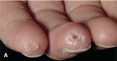
Scleroderma (lSSc): acrosclerosis (A) Typical "rat bite" necroses and ulcerations of fingertips. Fitzpatrick Figure 14-46
answer
Identify this condition
question: acrosclerosis (B) Thinning of lips—microstomia (which would show better when patient attempts to open her mouth), radial perioral furrowing. Beaklike sharp nose. Fitzpatrick Figure 14-46")
Scleroderma (lSSc): acrosclerosis (B) Thinning of lips—microstomia (which would show better when patient attempts to open her mouth), radial perioral furrowing. Beaklike sharp nose. Fitzpatrick Figure 14-46
answer
Identify
question
Scleroderma (dSSc) Mask-like facies with stretched, shiny skin and loss of normal facial lines giving a younger appearance than actual age; the hair is dyed. Thinning of the lips and perioral sclerosis result in a small mouth. Sclerosis (whitish, glistening areas) and multiple telangiectases (not visible at this magnification) are also present. Fitzpatrick Figure 14-47
answer
Describe this condition
question
Scleroderma: CREST syndrome Fitzpatrick Figure 14-48
answer
What is this condition associated with?
question
Scleroderma: CREST syndrome Numerous macular or matlike telangiectases on the forehead. Complete features include calcinosis cutis, Raynaud phenomenon, esophageal dysmotility, sclerosis, and telangiectasia. Fitzpatrick Figure 14-48
answer
Identify
question
Sarcoptes scabiei—lesions. Note three arrows that point to linear track-like lesions on the hand. (Reproduced with permission from Wolff K, Johnson R. Fitzpatrick's Color Atlas & Synopsis of Clinical Dermatology. 6th ed.
answer
Identify
question
Bacteria Spirochetes: Borrelia burgdorfer (Toronto notes 2014)
answer
What causes lyme disease
question
serology: ELISA, Western Blot (Toronto notes 2014)
answer
Lyme disease diagnostics are...?
question
stage 1: doxycycline amoxicillin cefuroxime stage 2-3: ceftriaxone (Toronto notes 2014)
answer
Lyme disease treatment
question
Acute urticaria and angioedema Note that there are both superficial wheals and deep, diffuse edema. Occurred after the patient had eaten shellfish. He had similar episodes previously but never considered seafood as the cause. Fitzpatrick Figure 14-7
answer
Identify
question
Cholinergic urticaria Small urticarial papules on neck occurring within 30 min of vigorous exercise. Papular urticarial lesions are best seen under side lighting. Fitzpatrick Figure 14-10
answer
Identify
question
Urticaria: dermographism Urticaria as it appeared 5 min after the patient was scratched on the back. The patient had experienced generalized pruritus for several months with no spontaneously occurring urticaria. Fitzpatrick Figure 14-09
answer
Identify
question
Acute urticaria 1. Antihistamines: H1-blockers, e.g., hydroxyzine, terfenadine; or loratadine, cetirizine, fexofenadine; 2. Prednisone 3. Danazol or Stanozolol Fitzpatrick's Figure 14-6
answer
What is the treatment for this condition?
question
Chronic urticaria Chronic urticaria of 5-year duration in an otherwise healthy 35-year-old female. Eruptions occur on an almost daily basis and, as they are highly pruritic, greatly impair the patient's quality of life. Although suppressed by antihistamines, there is an immediate recurrence after treatment is stopped. Repeated laboratory and clinical examinations have not revealed an apparent cause. Fitzpatrick's Figure 14-8
answer
Identify
question
Urticaria: dermographism Autoimmune urticaria is tested by the autologous serum skin test and determination of anti-FcϵRI antibody. If urticarial wheals do not disappear in ≤24 h, urticarial vasculitis should be suspected and a biopsy done. Fitzpatrick's Figure 14-9
answer
How is this condition diagnosed?
question
Tinea Capitis: Dermatophyte infection To establish a diagnosis, hair shafts should be plugged out and cultured, as well as examined after KOH preparation. Fitzpatrick's Figure 88-22
answer
How is a diagnosis established for this condition?
question
Tinea Capitis: Dermatophyte infection Trichophyton tonsurans accounts for around 90% of cases of tinea capitis in the United States and Unites Kingdom Fitzpatrick's Figure 88-22
answer
What causes this condition?
question
Tinea Capitis: Dermatophyte infection The alopecic patches usually show signs of inflammation and scaling with brittle grayish hair stumps Fitzpatrick's Figure 88-22
answer
Describe this condtion
question
Tinea Capitis: Dermatophyte infection The alopecic patches usually show signs of inflammation and scaling with brittle grayish hair stumps.The areas may show a yellow-green fluorescence under Wood's Light examination. Fitzpatrick's Figure 88-22
answer
What test is used to analyze the alopcic patches? And what is seen?
question
Tinea Capitis: Dermatophyte infection Systemic antifungal treatment is indispensable to treat tinea capitis. Topical sporicidal agents, such as selenium or ketoconazole help to limit the spread of the infectious spores, but cannot be used without systemic antifungal treatment. Fitzpatrick's Figure 88-22
answer
What is first line treatment for this condition?
question
Acanthosis nigricans Acanthosis nigricans involving the axilla with numerous acrochordons. Fitzpatrick's Figure 153
answer
Identify
question
Acanthosis nigricans The clinical hallmark of acanthosis nigricans is development of gray-brown, velvety plaques that may start as a dirty appearance. Fitzpatrick's Figure 153
answer
What is the hallmark of this condition?
question")
Acanthosis nigricans Acanthosis nigricans: typical hyperpigmented, velvety, verrucous axillary plaques. (Reproduced, with permission, from Wolff K, Johnson RA, Suurmond D. Fitzpatrick's Color Atlas & Synopsis of Clinical Dermatology. 6th ed. New York: McGraw-Hill; 2005. Fig. 5-1.)
answer
Identify
question
Acanthosis nigricans The most commonly involved locations are the axillae, neck, external genitalia, groin, face, inner thighs, antecubital and popliteal fossae, umbilicus, and perianal area. Acrochordons may develop, superimposed on the acanthosis nigricans or on other locations Fitzpatrick's Figure 153
answer
What are the most common sites for this condition?
question
Acanthosis nigricans... A. Verrucous and papillomatous growths of the vermillion border of the lip. Fitzpatrick's Figure 153-2
answer
The growths seen in this picture are associated with what condition?
question
Acanthosis nigricans... B. Velvety thickening of the tongue. Fitzpatrick's Figure 153-2
answer
The thickening of the tongue in this picture is associated with what condition?
question
Malignant acanthosis nigricans Concerns for malignancy-associated acanthosis nigricans should arise when a rapid appearance of the lesions in an older individual along with atypical sites such as the oral mucosa is involved Fitzpatrick's Figure 153-2
answer
When someone is diagnosed with __________ _________ and sites noted in the picture are involved, what is of concern?
question
Acanthosis nigricans. Although the precise etiology of benign acanthosis nigricans remains unclear, there is evidence that insulin plays a significant role (see Chapter 151) Fitzpatrick's Figure 153
answer
What is the cause of this condition?
question
Malignant acanthosis nigricans Malignant acanthosis nigricans typically occurs in older patients and frequently coexists with other paraneoplastic dermatoses such as tripe palms and the sign of Leser-Trélat. Fitzpatrick's Figure 153-2
answer
What population is more at risk for the condition that involves these atypical sites (oral mucosa)?
question
Acanthosis nigricans The lesions of malignant and benign acanthosis nigricans are indistinguishable. Fitzpatrick's Figure 153
answer
How are malignant and benign acanthosis nigricans distinguished?
question
Fitzpatrick's Figure 153 Management of any co-occurring disease or malignancy often improves and may even resolve acanthosis nigricans. Topical keratolytics, including the retinoids, and oral retinoids can reduce the appearance of acanthosis nigricans. Other oral medications reported to show improvement include dietary fish oil, metformin, and cyproheptadine, possibly by inhibition of tumor secreted growth factors in the case of the latter. Fitzpatrick's Figure 153
answer
How is this condition treated?
question
Melasma Well-demarcated, hyperpigmented macules are seen on the cheek, nose, and upper lip. Fitzpatrick's Figure 13-10
answer
Identify
question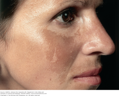
Melasma Management: hydroquinone 3% solution and 4% cream; azelaic acid 20% cream; and a combination of fluocinolone 0.01%, hydroquinone 4%, and tretinoin 0.05%. Hydroquinone 4% cream can be compounded with 0.05% tretinoin cream or glycolic acid by the pharmacist. Prevention: Opaque sun blocks. Synonyms: Chloasma (Greek: "a green spot"), mask of pregnancy. Fitzpatrick's Figure 13-10
answer
What are the treatment options for this condition?
question
It may be associated with pregnancy, with ingestion of contraceptive hormones, or possibly with certain medications such as diphenylhydantoin, or it may be idiopathic. Very common, especially among persons with constitutive brown skin taking contraceptive pills and living in sunny climates; 10% of patients are men. Fitzpatrick's Figure 13-10
answer
What is this condition associated with?
question
Vitiligo Vitiligo: face Extensive depigmentation of the central face. Involved vitiliginous skin has convex borders, extending into the normal pigmented skin. Note the chalk-white color and sharp margination. Note also that the dermal nevomelanocytic nevus on the upper lip has retained its pigmentation. Fitzpatrick's Figure 13-2
answer
Identify
question
Vitiligo 1. Inheritance 2. Family history of thyroid disease, diabetes mellitus, and vitiligo appear to be at increased risk for development of vitiligo. 3. Many patients attribute the onset of their vitiligo to physical trauma (where vitiligo appears at the site of trauma—Koebner phenomenon), illness, or emotional stress. Onset after the death of a relative or after severe physical injury is often mentioned. A sunburn reaction may precipitate vitiligo. Fitzpatrick's Figure 13-3
answer
What are the possible causes for this condition?
question
Three principal theories have been presented about the mechanism of destruction of melanocytes in vitiligo: 1. The autoimmune theory holds that selected melanocytes are destroyed by certain lymphocytes that have somehow been activated. 2. The neurogenic hypothesis is based on an interaction of the melanocytes and nerve cells. 3. The self-destruct hypothesis suggests that melanocytes are destroyed by toxic substances formed as part of normal melanin biosynthesis. Fitzpatrick's Figure 13-2
answer
What are the 3 possible ways melanocytes are destroyed in this condition?
question
Universal vitiligo Vitiliginous macules have coalesced to involve all skin sites with complete depigmentation of skin and hair in a female. The patient is wearing a black wig and has darkened the brows with eyebrow pencil and eyelid margins with eye liner. Fitzpatrick's Figure 13-5
answer
Identify
question
Vitiligo: predilection sites. Fitzpatrick's Figure 13-4
answer
FYI Vitiligo: predilection sites.
question
Vitiligo: Treatment: Excimer laser (308 nm) Best results in the face. Topical glucocorticoids: Monitor for signs of early steroid atrophy. Topical calcineurin inhibitors: Tacrolimus and pimecrolimus. They are reported to be most effective when combined with UVB or excimer laser therapy. Topical photochemotherapy [topical 8-methoxypsoralen (8-MOP) and UVA] Fitzpatrick's Figure 13-2
answer
What are the treatment options for this condition?
question
Generalized Vitiligo Systemic photochemotherapy: Narrow-band UVB, 311 nm: This is just as effective as PUVA and does not require psoralens. It is the treatment of choice in children <6 years of age. Fitzpatrick's Figure 13-3
<img src="https://chmanchacentro.com/wp-content/uploads/2018/04/generalized-vitiligosystemic-photochemotherapy-narrow-band-uvb-311-nm-this-is-just-as-effective-as-puva-and-does-not-require-psoralens-it-is-the-treatment-of-choice-in-children.png" title="Generalized Vitiligo Systemic photochemotherapy: Narrow-band UVB, 311 nm: This is just as effective as PUVA and does not require psoralens. It is the treatment of choice in children <6 years of age. Fitzpatrick's Figure 13-3" alt="Generalized Vitiligo Systemic photochemotherapy: Narrow-band UVB, 311 nm: This is just as effective as PUVA and does not require psoralens. It is the treatment of choice in children
answer
What is the treatment option for the "generalized" version of this condition?
question
Vitiligo Topical calcineurin inhibitors: Tacrolimus and pimecrolimus. They are reported to be most effective when combined with UVB or excimer laser therapy. Fitzpatrick's Figure 13-2
answer
What is the MOST EFFECTIVE topical treatment for this condition, when combined with laser therapy?
question
Psoriasis A-F. Chronic plaque psoriasis located at typical sites. Note marked symmetry of lesions. Cutaneous Lesions: The classic lesion of psoriasis is a well-demarcated, raised, red plaque with a white scaly surface (Fig. 18-7). Lesions can vary in size from pinpoint papules to plaques that cover large areas of the body. Fitzpatrick's Figure 18-7
answer
Identify
question
Psoriasis A-F. Chronic plaque psoriasis Cutaneous Lesions: The classic lesion of psoriasis is a well-demarcated, raised, red plaque with a white scaly surface (Fig. 18-7). Lesions can vary in size from pinpoint papules to plaques that cover large areas of the body. Fitzpatrick's Figure 18-7
answer
Describe the classical lesion of this condition
question
Psoriasis Auspitz sign. Note point of bleeding after scale is removed. Under the scale, the skin has a glossy homogeneous erythema, and bleeding points appear when the scale is removed, traumatizing the dilated capillaries below (the Auspitz sign) (Fig. 18-8). Fitzpatrick's Figure 18-8
answer
The appearance of punctate bleeding spots in this condition is called what?
question
Psoriasis Vulgaris Psoriasis vulgaris is the most common form of psoriasis, seen in approximately 90% of patients. Fitzpatrick's Figure 18-7
answer
What is the most common type of this condition?
question, hands (B), and back (C and D). The patient in D went on to develop chronic plaque psoriasis. Fitzpatrick's Figure 18-11")
Guttate psoriasis Guttate psoriasis, involving thigh (A), hands (B), and back (C and D). The patient in D went on to develop chronic plaque psoriasis. Fitzpatrick's Figure 18-11
answer
Identify
question
Erythrodermic psoriasis. Erythrodermic psoriasis: The patient shown in panel A rapidly developed near-complete involvement and complained of fatigue and malaise. Note islands of sparing. Fitzpatrick's Figure 18-13
answer
Identify
question
Erythrodermic psoriasis. The patient shown in panels B and C had total body involvement with marked hyperkeratosis and desquamation. Fitzpatrick's Figure 18-13
answer
Identify
question
Nail psoriasis. Panel C demonstrates subungual hyperkeratosis. Fitzpatrick's Figure 18-15
answer
Identify
question
Nail psoriasis. Panel B demonstrates nail pitting. Fitzpatrick's Figure 18-15
answer
Identify
question
Nail psoriasis. Panel A demonstrates distal onycholysis and oil drop spotting. Fitzpatrick's Figure 18-15
answer
Identify
question
Nail psoriasis. Panel D demonstrates onychodystrophy and loss of nails in a patient with psoriatic arthritis. Fitzpatrick's Figure 18-15
answer
Identify
question
Chronic plaque psoriasis Methotrexate MTX is highly effective for chronic plaque psoriasis and is also indicated for the long-term management of severe forms of psoriasis, including psoriatic erythroderma and pustular psoriasis. Fitzpatrick's Figure 18-7
answer
What are the treatment options for this SPECIFIC condition?
question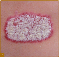
Psoriasis Corticosteroids Vitamin D3 and Analogs Anthralin (Dithranol) Coal Tar Tazarotene Topical Calcineurin Inhibitors And many more Fitzpatrick's Figure 18-7 CH 18 "Treatment" section
answer
What are the treatment options for this condition?
question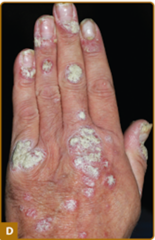
Chronic plaque psoriasis There is no known prevention for psoriasis. CH 18 "Prevention" section
answer
How is this condition prevented?
question
Pressure ulcers Common sites of pressure ulcer development. Figure 100-2
answer
Just FYI
question
Pressure ulcer Pressure ulcer, stage III, complicated by fecal incontinence. Fitzpatrick's Figure 100-2
answer
Identify
question Thrush is a yeast infection from Candida species. Shown here on the palate, it may appear elsewhere in the mouth (see p. 289). Thick, white plaques are somewhat adherent to the underlying mucosa. Bates pg 285")
Thrush on the Palate (Candidiasis) Thrush is a yeast infection from Candida species. Shown here on the palate, it may appear elsewhere in the mouth (see p. 289). Thick, white plaques are somewhat adherent to the underlying mucosa. Bates pg 285
answer
Identify
question Predisposing factors include (1) prolonged treatment with antibiotics or corticosteroids and (2) AIDS. Bates pg 285")
Thrush on the Palate (Candidiasis) Predisposing factors include (1) prolonged treatment with antibiotics or corticosteroids and (2) AIDS. Bates pg 285
answer
What are the predisposing factors for this condition?
question
Candidiasis. Note the thick white coating from Candida infection. The raw red surface is where the coat was scraped off. Infection may also occur without the white coating. Bates pg 289
answer
Identify
question
Candidiasis It is seen in immunosuppression from chemotherapy or prednisone therapy. Bates pg 289
answer
Who are at risk for this condition
question
Molluscum Contagiosum Dome-shaped, fleshy lesions Bates pg 880
answer
Identify
question and occipital lymph node (arrow). Bates pg 880")
Tinea Capitis Scaling, crusting, and hair loss are seen in the scalp, along with a painful plaque (kerion) and occipital lymph node (arrow). Bates pg 880
answer
Identify
question in a drug eruption in an infant Bates pg 197")
Urticaria Wheals (urticaria) in a drug eruption in an infant Bates pg 197
answer
Identify
question
Kaposi's sarcoma in AIDS: This malignant tumor may appear in many forms: macules, papules, plaques, or nodules almost anywhere on the body. Lesions are often multiple and may involve internal structures. On left: ovoid, pinkish red plaques that typically lengthen along the skin line may become pigmented. On right: a purplish red nodule on the foot. Bates pg 197
answer
Identify
question
Kaposi's sarcoma: Several chemotherapeutic regimens For patients with anthracycline-refractory AIDS KS paclitaxel Interferon-α, which has been a mainstay therapeutic approach for AIDS KS during the 1980s and early 1990s, still holds some promise for AIDS patients with early disseminated KS who simultaneously receive HAART. Fitzpatrick's Figure 128-1
answer
What are the treatment options
question
Kaposi's sarcoma: Classic variant. Plaques and papules localized on the dorsum of the foot, a site of predilection of classic KS. Fitzpatrick's Figure 128-1
answer
Identify
question
Kaposi's sarcoma: Tumor nodules of advanced classic KS with severe involvement of extremities. Fitzpatrick's Figure 128-2
answer
Identify
question
Kaposi's sarcoma: KS lesions on the hard palate are typical manifestations of AIDS-associated KS. Fitzpatrick's Figure 128-4
answer
Identify
question
Psoriasis Bates pg 197
answer
Identify
question
Tinea Captits ("Ringworm") Round scaling patches of alopecia. Hairs are broken off close to the surface of the scalp. Usually caused by fungal infection from Trichophyton tonsurans from humans, less commonly from microsporum canis from dogs or cats. Bates pg 201
answer
Identify
question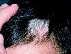
Tinea Captits ("Ringworm") Mimics seborrheic dermatitis. Bates pg 201
answer
What other condition does the one in this picture mimic?
question
Crusted scabies. Hyperkeratotic plaques populated with thousands of mites. Fitzpatrick's 8e Figure 208-1
answer
Identify
question
Molluscum contagiosum Molluscum contagiosum infection. A patient with advanced HIV/AIDS presented with multiple large facial mollusca causing significant cosmetic disfigurement. Fitzpatrick's 8e Figure 198-13
answer
Identify
question
Scabies Scabies. Several thread-like burrows are present in the web spaces of the fingers and on the knuckles, a common location for these lesions in scabies. Longitudinal scraping of a burrow will often reveal the mite or mite products under microscopic examination. Fitzpatrick's 8e Figure 208-2
answer
Identify
question
Crusted scabies Crusted scabies. Close-up showing erosions, lakes of pus, scales, and crusts. Figure 208-3.2
answer
Identify
question
Scabies The itching experienced during this time period is commonly referred to as "postscabetic itch." Fitzpatrick's 8e Figure 208-2
answer
What is another term for the itching/pruritis associated with this condition?
question
Lice: Pediculosis pubis Pediculosis pubis. Several lice and their dot-like nits attached to the hair shafts can be seen in the pubic area of this patient. Fitzpatrick's 8e Figure 208-6
answer
Identify
question Fitzpatrick's 8e Figure 208-7")
Lice Pediculosis pubis. Eyelash infestation with Pthirus pubis. Nits can be seen attached to the eyelashes. (Used with permission from D.A. Burns, MD.) Fitzpatrick's 8e Figure 208-7
answer
Identify
question
Lice (crab) Shaving is not curative as the louse will seek another hairy area of the body to reside. Crab lice are treated with the same topical therapy as that for pediculosis capitis (Box 208-4). Permethrin 1%a (Nix) Permethrin 5% Malathion 0.5% Carbaryl 0.5% Lindane 1% Topical ivermectin Ivermectin, oral 200 μg/kg Fitzpatrick's 8e Box 208-4 Treatment of Head Lice and Crab Lice
answer
How is this condition treated?
question
Staphylococcus aureus: bullous impetigo. Local treatment with mupirocin ointment or cream, removal of crusts, and good hygiene is sufficient to cure most mild to moderate cases. Retapamulin 1% ointment Fusidic acid Systemic antibiotics may be required in extensive cases. Fitzpatrick 8e Figure 176-8
answer
How is this condition treated?
question
Furuncle Simple furunculosis may be aided by local application of moist heat. A carbuncle or a furuncle with surrounding cellulitis, or one with associated fever, should be treated with a systemic antibiotic (as for MRSA impetigo; see Box 176-4). Fitzpatrick 8e Figure 176-8
answer
This condition with surrounding cellulitis is treated with what?
question
Multiple furuncles B. Multiple furuncles. Multiple abscesses on the buttocks of long standing in a young man with inflammatory bowel disease. The lesions healed with scarring after a prolonged course of dicloxacillin Vancomycin (1.0-2.0 g intravenously daily in divided doses) or other systemic parenteral agents that have anti-CA-MRSA activity are indicated for these patients. Antibiotic treatment should be continued for at least 1 week. Fitzpatrick 8e Figure 176-8
answer
What is the treatment for this condition
question
Staphylococcus aureus whitlow (felon). A pyogenic granuloma arose 1 week after trauma to the bulb of the thumb. A week later, the bulb became swollen, erythematous, and very tender. Abscess formation is seen with loculation of pus. X-ray films showed early osteomyelitis complicating the whitlow. Fitzpatrick's FIGURE 176-13
answer
Identify
question
Three types of skin eruptions can be produced by phage group II S. aureus, particularly strains and: (1) bullous impetigo, (2) exfoliative disease (SSSS), and (3) nonstreptococcal scarlatiniform eruption (staphylococcal scarlet fever). All three represent varying cutaneous responses to extracellular exfoliative toxins ("exfoliatin") types A and B produced by these Staphylococci (see Chapter 177). Fitzpatrick's CH 176
answer
What are the 3 eruption produced by S. aureus phage group II?
question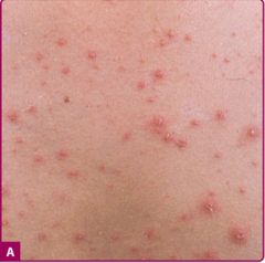
Varicella. A. A full spectrum of lesions—that is, erythematous papules, vesicles ("dewdrops on rose petals"), crusts, and erosions at sites of excoriation—is seen in a child with a typical case of varicella. Fitzpatrick's 8e FIGURE 194-3
answer
Identify
question
Varicella: In normal children, varicella is generally benign and self-limited. Cool compresses or calamine lotion locally, tepid baths with baking soda or colloidal oatmeal (three cups per tub of water) and oral antihistamines may relieve itching. The nucleoside analogues acyclovir, famciclovir, valacyclovir, and brivudin and the pyrophosphate analog foscarnet show efficacy in treating VZV infections. Fitzpatrick's 8e FIGURE 194-3
answer
What are the treatment options
question
Varicella. B. A wider range of lesions, including many large pustules, is seen in a 21-year-old female who was febrile as well as "toxic" and had varicella pneumonitis. Fitzpatrick's 8e FIGURE 194-3
answer
Identify
question
Erysipelas. Painful, edematous erythema with sharp margination on both cheeks and the nose. There is tenderness, and the patient has fever and chills. Fitzpatricks 8e Figure 178-4
answer
Identify
question")
Necrotizing fasciitis Treatment of all necrotizing SSTI involves a combination of urgent debridement, antibiotics, and often, adjunctive therapies. (Fitzpatricks 8e Figure 179-1)
answer
How is this condition treated?
question with progressive necrosis of the pubic, perigenital, and perianal tissue. (Fitzpatricks 8e Figure 179-1)")
Necrotizing fasciitis Fournier's gangrene (type 1 necrotizing fasciitis of genitalia) with progressive necrosis of the pubic, perigenital, and perianal tissue. (Fitzpatricks 8e Figure 179-1)
answer
Identify

