Veterinary Neurology – Flashcards
Unlock all answers in this set
Unlock answersquestion
Discospondylitis
answer
Inflammation due to infection of vertebral articulations
question
Intervertebral Disc Disease
answer
Herniation/prolapse of disc material into spinal canal
question
canine vertebral formula
answer
C-7, T-13, L-7, S-3
question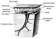
What is the function of the meninges?

answer
Provide physical protection & support Allow the carriage of blood through blood vessels Contains the CSF
question
Pia mater
answer
Innermost membrane surrounding the brain and spinal chord containing a network of blood vessels that nourish the nervous tissue
question
arachnoid
answer
Middle membrane surrounding the brain and spinal chord - looks like a spider web
question
dura mater
answer
Surrounding the whole brain and spinal chord is the strongest layer
question
Subarachnoid space
answer
The pia mater and the arachnoid make up the space wherein flows part of the cerebral spinal fluid exists
question
Where is the sub dural space?
answer
Space between the Dura and the arachnoid
question
Falx cerebri
answer
found in the longitudinal fissure between the cerebral hemispheres -- divides the brain in to left and right hemispheres
question
Tentorum cerebelli
answer
separates the cerebrum from the cerebellum
question
Describe the spinal cord basic anatomy.
answer
Grey matter is in the darker portion, white matter is surrounding it
question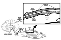
Ventral Grey Matter Horn

answer
Motor movement is controlled there
question
Ventral root
answer
contains axons of somatic motor neurons and sometimes visceral motor neurons that control peripheral effectors; each segment of the spinal cord is associated with a pair of these
question
Spinal Cord
answer
A column-shaped continuation of the brainstem (medulla), extending from the foramen magnum (base of the skull) to the lumbar region of the vertebral column. Composed of centrally-located gray matter and the surrounding white matter
question
Lower motor neuron
answer
Lives in the ventral grey matter horn, leaves through the ventral root second order- originates in brainstem or spinal cord (cranial nerve nuclei or ventral horn of spinal cord)-supply effectors (muscles) to stimulate movement-axon of this motor neuron leads the rest of the way to the muscle or other target organ- damage produces spastic paralysis
question
Dorsal Root
answer
Sensory neurons come in to the spinal cord through this root. (receives information regarding touch and sensation)
question
Gray Matter of the Spinal Cord
answer
A butterfly-shaped region surrounding the central canal, it consists primarily of neuron (nerve) cell bodies
question
Dorsal Root Ganglion
answer
located on dorsal root, cell bodies of the afferent nerves (somatic and visceral sympathetic)
question
White Matter of the Spinal Cord
answer
The outer region of the spinal cord, mostly composed of myelinated and unmyelinated nerve fibres (axons) that carry impulses to and from the brain. Its regional divisions (dorsal, lateral and ventral) are called columns, or tracts, in which specific types of information are relayed along the spinal cord, e.g. sensory vs motor. Contains mostly axons and dendrites.
question
If you damage the spinal chord at C7, will this affect reflexes in the thoracic limb?
answer
Yes, there were be damage at that segmental level specifically - so reflexes will be off
question
Spinal reflex
answer
reflex in which important interconnections and processing occur inside the spinal cord; range from monosynaptic to polysynaptic
question
White Matter of the Spinal Cord
answer
The outer region of the spinal cord, mostly composed of myelinated and unmyelinated nerve fibres (axons) that carry impulses to and from the brain. Its regional divisions (dorsal, lateral and ventral) are called columns, or tracts, in which specific types of information are relayed along the spinal cord, e.g. sensory vs motor.
question
Spinal Cord Enlargements
answer
There are two regional enlargements of the spinal cord for the innervation of the limbs: · Cervical : composed of cord sections C6, C7, C8, T1, (T2). · Lumbosacral : composed of cord sections L5, L6, L7, S1 (Enlargement L4 to S3)
question
Dorsal Root Ganglia
answer
A swelling located at the entrypoint into the spinal cord of the dorsal root of the spinal nerve, containing the cell bodies of afferent somatic sensory neurons, i.e. neurons concerned with sensing touch, pressure, vibrations, temperature, pain, and myotactic positioning (e.g. Golgi Tendon Organ receptors).
question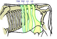
Dermatome

answer
circumscribed skin area that is supplied mainly from one spinal cord segment through a particular spinal nerve
question
Mixed Spinal Nerve
answer
where the ventral and dorsal roots pass through the intervertebral foramen and come in close proximity to one another. Contains both motor and sensory nerve fibers
question
Cerebral Spinal Fluid
answer
* A fluid produced by the choroid plexus within the ventricular system of the brain. * CSF is characterized by having low protein, few cells, and a low number of ions
question
What makes white matter?
answer
Astrocytes
question
What makes grey matter?
answer
Primarily Schwann cells
question
How is CSF characterized?
answer
having low protein, few cells, and a low number of ions.
question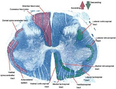
Spinal Tracts

answer
The white columns of the spinal cord that provide two-way conduction paths to and from the brain; ascending tract carries information to the brain, whereas descending tracts conduct impulses from the brain
question
How do you check for reflex response in areas of the trunk which are not serviced by an enargement area?
answer
Use a pinprick sensation test to see when the animal reacts. This is called a dermatome
question
Dermatome
answer
circumscribed skin area that is supplied mainly from one spinal cord segment through a particular spinal nerve
question
Choroid Plexus
answer
capillary-rich folds of membrane in the ventricles, covered by a thin cuboidal membrane. produces CSF - squeezes out fluid
question
What are the two places in the spinal cord which are action sensitive? (high traffic?)
answer
The places (enlargements) which innervate the hind legs (Lumbosacral) and the front legs(Cervicalthoracic). These areas are larger because there is more grey matter.
question
What sections are the cervicalthoracic enlargement composed of?
answer
composed of cord sections C6, C7, C8, T1, (T2) (know the ones in the brackets as well)
question
What sections are the lumbosacral enlargement compsed of?
answer
composed of cord sections L5, L6, L7, S1 (some say L4 to S3)
question
Intervertebral disc disease
answer
Degenerative changes increase with repetitive compression (e.g. heavy lifting in flexion) or trauma (e.g. fall); degenerative changes may be asymptomatic
question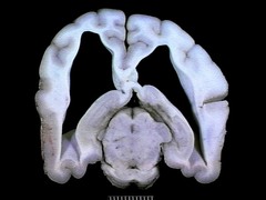
Non-communicating Hydrocephalus

answer
increase in CSF volume and pressure due to obstructed CSF flow within the ventricular system (most often at the cerebral aqueduct). Pressure increases inside the ventricular system relative to the subarachnoid space. - rapidly developing
question
Communicating Hydrocephalus
answer
similar to non-communicating, BUT due to problems with CSF uptake at arachnoid granulations. Pressure increased outside and inside ventricular system. - slowly developing
question
Arachnoid Granulations
answer
specialized regions of the arachnoid containing microscopic tubules and venules with modified endothelium to allow passive uptake of CSF.
question
What is the CSF circulatory flow pattern?
answer
1. Choroid Plexus 2. Cerebral Aqueduct 3. Fourth Ventricle 4. Foramina of Luska 5. Subarachnoid Space (between arachnoid & pia) 6. Arachnoid Granulations 7. Dural Venous Sinus (pooled aggregates of veins, reabsorption) a) Dorsal Sagittal Venous Sinus b) Transverse Sinus - in the Tentorium Cerebelli. 8. Blood Stream
question
Cauda Equina
answer
bunch of nerve fibers at the end of the spinal cord that appear like a horse's tail
question
Why do some come off prior to the disc and sometimes after the disc?
answer
The discrepancy at C8
question
Ventral spinal artery
answer
Follows ventral surface of cord and provides blood supply to the spinal cord
question
Paired dorsolateral spinal arteries
answer
runs along base of dorsal roots of spinal nerves and provides blood supply to the spinal cord
question
How is blood supply delivered to the spinal cord?
answer
Segmentally supplied by branches of the vertebral artery (in cervical and anterior thoracic regions), the intercostal artery (in thoracic region), and the aorta (in lumbar region) that feed into three arteries that run the length of the cord:
question
How do reflex transmissions get into the spinal cord?
answer
If information is getting on to and off of the spinal cord 'highway' at the same spot, then they don't use tracts
question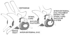
Fibrocartilaginous Embolic Myelopathy (FCEM)

answer
fibrocartilaginous emboli plug a radicular artery, resulting in the vascular embarrassment of a segment of cord.
question
Somatotopic
answer
different parts of a given region associated with distinct parts of the body, both functionally and anatomically.
question
Contralateral
answer
on or relating to the opposite side (of the body)
question
Decussates
answer
cross from one side of the CNS to the other
question
Where is the primary neuron's (nerve) cell body ALWAYS found?
answer
Dorsal Root Ganglion
question
Where does the primary neuron ALWAYS enter the spinal cord?
answer
The dorsal root
question
IPSILATERAL
answer
on or relating to the same side (of the body)
question
What is the exception where the axon enters the spinal cord and ascends on the same side?
answer
unconscious proprioception
question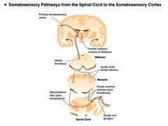
Conscious Sensation

answer
awareness -preceived by cerebral cortex - exteroceptive - proprioceptive
question
Unconscious Proprioception
answer
Hind Limbs: dorsal spinocerebellar and ventral spinocerebellar tracts Front Limbs: Rostral spinocerebellar tracts
question
Proprioception
answer
the ability to sense the position and location and orientation and movement of the body and its parts
question
The Touch/Pressure tracts are carried in the same tracts as what?
answer
Proprioception (i.e. Fasciculus Gracilis) Note that very light touch is in the Spinothalamic tracts
question
Dorsal Column
answer
Located at the most dorsal aspect of the spinal cord, it includes the medial Fasciculus Gracilis (hindlimbs, caudal to T6) and the more lateral Fasciculus Cuneatus (forelimbs, cranial to T6).
question
Fasciculus Cuneatus is a dorsal column which tend get sensory information (conscious proprioception - throacic limbs) from what region?
answer
Forelimbs, cranial to T6
question
Fasciculus Cuneatus
answer
(conscious proprioception) First-order sensory neurons contained in the dorsal funiculus that project to the nucleus cuneatus and that convey the modalities of conscious proprioception and tactile sensation from the ipsilateral side of the upper limb.
question
If you see an animal with conscious proprioception, what can you assume?
answer
Stumbling, issues with proprioception, doesn't react to knuckling response very quickly. Issues are found on both left and right sides (bilateral).
question
Fasciculus Gracilis
answer
(conscious proprioception - pelvic limbs) This tract extends to all levels of the spinal cord and contains fibers from the lower and upper limbs. Mediates conscious proprioception that include kinesthetics and discriminative touch (Generally - hindlimbs, caudal to T6)
question
Ventrolateral Tract
answer
Located in the ventrolateral areas of the cord's white matter. Carries pain and temperature sensations in fibres of moderate or small diameter and myelination.
question
What other problems will you see (besides a bilateral problem) when there is a problem in the spinal cord?
answer
Ataxia (hind end wobbley, scuffing of feet, tripping over its own feet)
question
Spinal Ataxia
answer
inability to coordinate voluntary muscle movements
question
If an animal has a problem in the spinal chord it will have:
answer
a) bilateral signs b) ataxia (can be subtle)
question
Ascending Reticular Activating System (RAS)
answer
afferent fibers running through the reticular formation that influence physiological arousal, without these you have continuous sleep
question
Central Nervous System
answer
Nervous tissue which lies within the brain and spinal cord.
question
Peripheral Nervous System
answer
Nervous tissue which lies outside the brain and spinal cord.
question
Peripheral Nerve
answer
A collection of axons within the peripheral nervous system which may carry afferent impulses (sensory nerve), efferent impulses (motor nerve) or both (mixed nerve). NB: A mixed nerve has axons which are sensory, and different axons which are motor.
question
Upper Motor Neuron
answer
Refers to all the neurons in the series from the brain outwards, except the final neuron which directly contacts the muscle. Any of the tracts from the brain which stay in the spinal cord (they can have synapses along the way)
question
Lower Motor Neuron
answer
The neuron in a motor pathway (somatic) which leaves the spinal cord and travels, via the ventral root, to the target muscle. (Can in some cases come out of the CNS)
question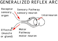
Reflexes

answer
A reflex is an automatic response to a stimulus (higher centres not required), e.g. for a spinal reflex arc, the decision to react is made at the level of the spinal cord, allowing an extremely rapid response without having to wait for input from the brain.
question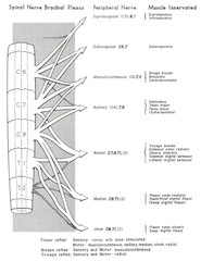
Brachial Plexus

answer
A pattern of organization of the peripheral nerves from spinal cord levels *C5 to T2* in which spinal nerves coalesce in a specific manner to allow innervation (sensory and motor) of specific regions/muscles by nerves from more than one spinal level
question
Lumbosacral Plexus
answer
A pattern of organization of the peripheral nerves from spinal cord levels *L4 to S2 * in which spinal nerves coalesce in a specific manner to allow innervation (sensory and motor) of specific regions/muscles by nerves from more than one spinal level
question
Radial Nerve
answer
largest branch of the brachial plexus, composed of C7, C8, T1
question
Sciatic Nerve
answer
arises from the sacral plexus and passes about halfway down the thigh where it divides into the common peroneal and tibial nerves (comprises L6, L7, S1)
question
Withdrawl Reflex
answer
Pinch the animal and look for the animal to withdraw its limb. Look for two things: 1. Withdrawl of the limb (multisynaptic relfex) 2. Reaction of the animal
question
Skeletomotor neuron (alpha motor neuron)
answer
* Innervates ordinary skeletal muscle, a.k.a. extrafusal motor fibres. * Often referred to as a motor neuron (motoneuron). * Has a large diameter axon and is therefore a fast conductor of APs. * Classed as an A alpha neuron and, as a result, is often referred to as an alpha motoneuron.
question
Fusimotor neuron (gamma motor neuron)
answer
* Innervates the intrafusal fibre which forms part of the muscle spindle. * Axon is thinner and slower conducting. * Classification as the A gamma neuron leads to it being referred to as the gamma motoneuron.
question
alpha motor neuron
answer
A lower motor neuron that goes to an extrafusal fibre and causes contraction
question
gamma motor neuron
answer
Goes to an extrafusal fibre resets the tension
question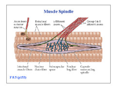
muscle spindle

answer
* a small striated muscle, the intrafusal muscle fibre, which has a stretchable middle portion (almost like a spring). * the annulospiral receptor, coils of sensory nerve endings wound around the central stretchable portion of the intrafusal fibre. * a fibrous capsule around the spindle that attaches indirectly to bone.
question
What does the muscle spindle detect?
answer
The muscle spindle detects stretch (of itself and its surrounding muscle body) by responding to an increase in muscle length - elongation of the stretchable central portion stimulates the coiled annulospiral receptor.
question
What is the first thing to do when a case comes in (and details of it)?
answer
Take a case history * Appetite * Attitude * Pain * Similar situation in the past
question
Corticospinal tract
answer
any of the important motor nerves on each side of the central nervous system that run from the sensorimotor areas of the cortex through the brainstem to motor neurons of the cranial nerve nuclei and the ventral horn of the spinal cord
question
Name 3 major descending pathways
answer
Rubrospinal tract Reticulospinal tract Vestibulospinal tract
question
Name 2 minor descending pathways
answer
Corticospinal tract Tectospinal tract
question
What are the enlargements that innervate the forlimbs, and what is it called?
answer
C5-T2, Cervicothoracic enlargement
question
Spinal segment
answer
region of cord from which a spinal nerve arises
question
Describe the recombination of the nerves that leave the spinal cord
answer
Although nerves leave from their named spinal segments, they will join neighbouring nerves to create a peripheral nerve that innervates a given muscle
question
Brachial Plexus
answer
a network of nerves formed by cervical and thoracic spinal nerves and supplying the arm and parts of the shoulder
question
Which spinal nerves contribute to the suprascapular nerve?
answer
(C5), 6, 7
question
Which spinal nerves contribute to the subscapular nerve?
answer
C6, C7
question
Which spinal nerves contribute to the musculocutaneous nerve?
answer
C6,7,8
question
Which spinal nerves contribute to the axillary nerve?
answer
(C6), 7, 8
question
Which spinal nerves contribute to the radial nerve?
answer
C7, C8, T1,(2)
question
Brachial plexus e____
answer
?? problem with nerves being stretched in limbs...
question
Which spinal nerves contribute to the median nerve?
answer
C8, T1, (2)
question
Which spinal nerves contribute to the ulnar nerve?
answer
C8, T1, (2)
question
Nerve roots are _____ to regenerate
answer
slow
question
What are the 3 events that happen after a sublethal injury of an axon?
answer
Degeneration Development of growth processes Re-establishment of contact (synapse) with target (muscle, other neuron)
question
Describe degeneration of an axon
answer
local anterograde (towards the axon terminal), and retrograde (towards the cell body)
question
What is development of new growth processes in a neuron that has been damaged due to?
answer
nerve growth factors and neurotrophins
question
Neurotrophins
answer
chemicals that promote neuron growth, required for the maintenance of sympathetic ganglia, and required for mature sensory neurons to regenerate after injury
question
Lower motor neuron
answer
second order- originates in brainstem or spinal cord (cranial nerve nuclei or anterior horn of spinal cord)-supply effectors (muscles) to stimulate movement-axon of this motor neuron leads the rest of the way to the muscle or other target organ- damage produces spastic paralysis
question
Transneuronal degeneration
answer
the intermediate neuron's death leads to adjacent neurons' deaths e.g. optic nerve
question
primary neuron
answer
also called first order neuron; functions as a sensory pathway neuron; originates in the root ganglia of spinal nerves; projects to the secondary neuron; the dendrites of this sensory neuron are part of the receptor that detects a specific stimulus.
question
reticular formation
answer
a nerve network in the brainstem that plays an important role in controlling arousal
question
Schiff-Sherrington response
answer
seen when the animal is in lateral recumbency. With damage to the propriospinal tract (the peri-gray white matter in the T3 to L3 spinal cord), the front leg exhibit extensor hypertonicity. The fore legs are not paralyzed; but, when left alone, will extend. The other component of the Schiff-Sherrington response is paralysis caudal to the lesion. This is due to the damage of the other white matter tracts. This is generally a sign of severe spinal cord damage, if both fore leg extensor hypertonicity and rear leg paralysis are seen, since the propriospinal tract is so deeply located within the white matter.
question
What segements of the spinal cord does the Femoral nerve come out of?
answer
L4, L5 L6 -; key spinal cord segment and nerve is L5 (Patellar reflex)
question
What segements of the spinal cord does the Sciatic nerve come out of?
answer
L6, L7, S1 (S2) -; key spinal nerve is L7 (flexor reflex)
question
What 4 tests do we use to evaluate the cotex?
answer
1. Menace response 2. Nasal septum response 3. Evaluating mentation a. thalamo-cortex - confusion, disorientation, changes in personality/behaviour b. brainstem - somnolence 4. Knuckling
question
Cortical ataxia
answer
Shows as some shaking and unsureness (some people debate that it doesn't exist)
question
polyneuropathy
answer
disease of many nerves (most often occurs as a side effect of diabetes mellitus, but may also occur as a result of drug therapy, critical illness such as sepsis, or carcinoma; exhibiting symptoms of weakness, distal sensory loss and burning)
question
Name 3 non-neurologic causes of knuckling
answer
1. Orthopedic disease • Metabolic • Hip dysplasia • Tendon rupture • Degenerative joint disease 2. Metabolic disease, e.g. weakness 3. Pain, e.g. fractures
question
What are the motor pathways
answer
...
question
Corticospinal
answer
direct control of movements below the head
question
Vestibulospinal
answer
transmits motor impulses that maintain muscle tone and activate ipsilateral limb and trunk extensor muscles and muscles that move head; in this way helps maintain balance during standing and moving
question
Reticulospinal
answer
descending tract whose fibers conduct motor impulses to sweat glands and muscles to control tone
question
Tectospinal
answer
reflexive orientating of head, trunk, eyes to visual or auditory stimulus
question
Rubrospinal
answer
origin: nucleus ruber (red nucleus) o influence foot musculature. funct:strongly influenced by cerebellum and cerebral cortex for muscle tone control primarily contralateral foot (all 4) musculature.
question
How is forelimb lameness characterized?
answer
head bobbing. The head is raised when the lame leg is on the ground and dropped when the sound limb is supporting the body.
question
Hypertrophic osteodystrophy
answer
seen in large and giant breed; young dogs in the rapid growth phase from 4 to 8 months · problems with the growing bones at the physis · extremely painful · not ataxic - predictable footfall, not floating in space
question
opisthotonus
answer
A type of spasm of the neck and back extensor muscles in which the head and heels arch backward in extreme hyperextension and the body forms a reverse bow.
question
How is hind-limb lameness characterized?
answer
by dropping of the forequarters as a result of overreaching by both forelimbs and by maintaining the head at a lower position than normal. Some breeds raise their tails when a lame hind limb is on the ground and drop it when the sound limb is on the ground.
question
Name the two main categories of proprioceptive fibres (ascending tracts) and the 2 major examples for each
answer
- Conscious Proprioception • Fasciculus cuneatus: thoracic limbs • Fasciculus gracilis: pelvic limbs - Reflex Proprioception • Rostral spinocerebellar tract: thoracic limbs • Dorsal & ventral spinocerebellar tract: pelvic limbs
question
Name the pathways that contain the nociceptive fibres.
answer
• Spinothalamic pathways (anterolateral system)
question
Spinothalamic
answer
passes up the anterior and lateral columns of the spinal cord, carries signals for main, temperature, pressure, tickle, itch, and light or crude (vaguely identifiable) touch - first order neurons end in the dorsal horn or the spinal cord, second order decussate to the opposite side of the spinal cord and there form the ascending spinothalamic tract and lead all the way to the thalamus - third order neurons continue from there to the cerebral cortex
question
What are signs that you have a lesion in C1-C5?
answer
UMN signs: 1. Spastic paresis/paralysis 2. Normal - hyperreflexia 3. Disuse atrophy (late & mild) 4. Normal to increased tone • Sensory loss signs:(same for all spinal regions) 1. Proprioceptive deficits/ataxia 2. Loss of nociception
question
What are signs that you have a lesion in C6-T2?
answer
LMN signs: 1. Flaccid paresis/paralysis 2. Hypo- to areflexia 3. Neurogenic atrophy (early & severe) 4. Hypo- to atonia • Sensory loss signs:(same for all spinal regions) 1. Proprioceptive deficits/ataxia 2. Loss of nociception
question
What are signs that you have a lesion in T3-L3?
answer
• UMN signs: 1. Spastic paresis/paralysis 2. Normal - hyperreflexia 3. Disuse atrophy (late & mild) 4. Normal - increased tone • Sensory loss signs: 1. Proprioceptive deficits/ataxia 2. Loss of nociception
question
What are signs that you have a lesion in L4-S3? (pelvic limbs and perineum)
answer
LMN signs: 1. Flaccid paresis/paralysis 2. Hypo- to areflexia 3. Neurogenic atrophy (early & severe) 4. Hypo- to atonia • Sensory loss signs:(same for all spinal regions) 1. Proprioceptive deficits/ataxia 2. Loss of nociception
question
Name 6 structures involved in spinal pain:
answer
- Bone ** - Nerve roots ** - Meninges ** - Ligaments - Articulations (synovial) - Disc annulus
question
What are 4 clinical signs of neck pain?
answer
• Stiff neck posture, low head carriage • Intermittent muscle spasms • Pain on vertebral palpation • Pain on vertical/horizontal flexion (don't do this if other signs present!) General Pain: - Increased respiratory ; heart rates - Pupillary dilation - Salivation - Pyrexia
question
What is the likely problem with a dog that - only wants to turn left - holding head to left - Subtle proprioceptive ataxia - whole body sway/ stagger (especially on turns) - Seemed almost "C" shaped - Spinal Reflexes: NAF
answer
NECK PAIN • C1-C5 left side white matter (ascending sensory tracts) • Final Diagnosis: Traumatic hemorrhage
question
Wobbler syndrome
answer
a condition of the cervical vertebrae that causes an unsteady (wobbly) gait and weakness (may include malformation of the vertebrae, intervertebral disc protrusion, and disease of the interspinal ligaments, ligamenta flava, and articular facets of the vertebra)
question
iatrogenic luxation
answer
A luxation due to a complication from a surgical procedure.
question
Calcinosis circumscripta
answer
tumoral condition of forming calcium deposit
question
What are your options for a further diagnostic work-up on a neurological case?
answer
• Electrodiagnostic studies - Electromyography (EMG) - Nerve conduction velocity studies (NCV) • Cerebrospinal fluid (CSF) collection ("spinal tap") ; analysis (cytology + protein) • Computed tomography (CT) / myelogram • Magnetic Resonance Imaging (MRI)
question
Neutrophilic pleocytosis
answer
An increase in neutrophilic cell count -; Steroid Responsive Meningitis
question
Anisocoria
answer
Unequal Pupil Size
question
What are signs of horner's syndrome?
answer
• Miosis (constriction of pupil) • Enophthalmos (sunken eyes) • Ptosis (drooping of the upper eyelid caused by muscle paralysis and weakness) • Protruding nictitans (Third eyelid) (T1, T2, T3 are affected)
question
Fibrocartilaginous Embolic Myelopathy
answer
-spinal cord infarction -fibrocartigalinous embolus obstructs ventral spinal artery -acute, non-progressive, nonpainful paralysis - Asymmetrical signs
question
What are some causes for mononeuropathies?
answer
- Neoplasia - Peripheral nerve sheath tumour (PNST) • Iatrogenic, e.g. injection into sciatic nerve • Inflammatory - Infectious vs immune-mediated • Trauma - Brachial plexus avulsion - Cauda equina syndrome - Tail avulsion (cat)
question
Brachial plexus avulsion
answer
Usually happens during a severe accident. The limb is wrenched from shoulder and the the dorsal and ventral roots can actually be pulled out of the spinal cord - lose motor and sensory sensation in limb -> paralyzed thoracic limb
question
Cauda Equine differentials (6)
answer
• Cauda Equina Syndrome - Lumbosacral disease • Anomaly / congenital • Intervertebral disc disease • Discospondylitis • Neoplasia • Trauma, e.g. fracture
question
Brachial plexus neuritis
answer
inflammation of the nerves of the brachial plexus accompanied by pain and sometimes loss of function (can be immune-mediated)
question
Causes of Polyneuropathies (4 major categories)
answer
• Immune-mediated / Idiopathic - Polyradiculoneuritis: "Coonhound Paralysis" - Vaccine-induced • Metabolic - Diabetes mellitus - Paraneoplastic, e.g. insulinoma - Hypothyroidism • Toxic: Organophosphates • Primary: Breed-related
question
Polyradiculoneuritis
answer
"Coonhound Paralysis" - Allergy to raccoon saliva. Epitope in raccoon saliva similar to epitope in ventral root and stimulates immune response • Acute onset pelvic limb weakness • Progressive to generalized weakness
question
diabetic polyneuropathy
answer
most common diabetic neuropathy, sensory symptoms usually occur before motor, sensory and motor changes distally and progress bilaterally to more proximal regions
question
Give some NMJ/Muscle: Differentials (5)
answer
• Myasthenia gravis • Acute polyradiculoneuritis (coonhound p.) • Bilateral anterior cruciate ligament rupture • Immune-mediate polyarthritis • Panosteitis
question
What are possible drugs used for treatment of myasthenia gravis?
answer
•Anticholinesterase drugs (congenital & acquired) - Pyridostigmine 1-3 mg/kg PO q.8-12 hrs - Neostigmine 2mg/kg/d IM • Immune-suppressive drugs (acquired form) - Prednisone (0.5mg/kg q.48hrs) - Azathioprine
question
Diencephalon
answer
This part/lobe of the brain is responsible for Body temperature regulation, pituitary hormone control, autonomic nervous system responses. Includes: thalamus, epithalamus, Hypothalamus (optic nerve comes out of here)
question
Pons
answer
part of the brain, works with the cerebellum in coordinating voluntary movement; neural stimulation studied in activation synthesis theory may originate here
question
Midbrain
answer
the middle division of brain responsible for hearing and sight; location where pain is registered; includes temporal lobe, occipital lobe, and most of the parietal lobe (CN III and IV come out of here)
question
What are the 3 main parts of the brainstem?
answer
Midbrain (sight, hearing, pain) Pons (Movement) - (CN Vm) Medulla (Breathing and heart function) - (CN VI to XII) (Also contains ARAS)
question
Vestibular system
answer
three semicircular canals that provide the sense of balance, located in the inner ear and connected to the brain by a nerve
question
What are the 4 degrees of modified mental status (neurological)?
answer
Alert Depressed Stuporous (Can be aroused with pain) Comatose
question
If a dog is disoriented, what are two potential causes?
answer
Thalamo-cortex Vestibular
question
Where do the cranial nerves originate?
answer
Diencephalon - CN II Midbrain - CN III IV Pons - CN Vm Medulla - CN VI - XII
question
Decerebrate rigidity
answer
results from damage to the upper brain stem (rostral-midbrain); all 4 limbs are hyperextended and spastic (poor prognosis)
question
opisthotonos
answer
state of a severe hyperextension and spasticity
question
Decerebellate rigidity
answer
Hind limbs not rigid or even flexed; mental status not affected; Cerebellar peduncles or rostral cerebellar lesion; (better prognosis)
question
Corneal Reflex
answer
shine a light toward a dog's eye. the light should be reflected in the same spot in the two corneas.
question
Otitis Media and Interna
answer
inflammation of the middle and inner ear structures. Can cause a facial nerve (CN VII) deficit
question
How does attributes of pathological nystagmus relate to the lesion?
answer
The quick phase is away from the lesion
question
What are 5 clinical signs of cerebellar problems?
answer
1. Cerebellar ataxia (Hypermetric/Wide-based) 2. Menace response deficits 3. Intention tremors (more in head than body) 4. Paradoxical vestibular signs 5. Others, e.g. decerebellate rigidity, anisocoria Note that there is *normal* mentation, postural reactions and strength
question
What are the two urinary sphincters, and what types of muscle are they made of?
answer
Internal - smooth (SNS - contraction - hypogastric- L1 - L4) External - skeletal (somatic - contraction - pudendal nerve S1 - S3)
question
Detrusor muscle
answer
formed by 3 layer of muscles contracts and compress urinary bladder to expel urine into urethra; (parasympathetic-contraction; sympathetic-relaxation)
question
Nerves involved in detrusor muscle innervation
answer
Hypogastric (L1 - L4) - filling - SNS Pelvic nerve (S1 - S3) - voiding - PSNS


