REFLUX ESOPHAGITIS, ESOPHAGEAL CANCER – Flashcards
Unlock all answers in this set
Unlock answersquestion
ESOPHAGEAL RINGS AND WEBS
answer
thin, typically delicate structures that partially or completely compromise the esophageal lumen. - present with dysphagia to solids
question
ESOPHAGEAL RING
answer
concentric, smooth, thin (axial length of 0.3-0.5 cm) extension of normal esophageal tissue consisting of 3 anatomic layers of mucosa, submucosa, and muscle - distal esophagus
question
A Ring
answer
uncommon, it is located approximately 1.5 cm proximal to the squamocolumnar junction, and it is rarely symptomatic.
question
B - Schatzki ring
answer
located at the squamocolumnar junction and are the most common type of esophageal ring
question
B - Schatzki ring
answer
almost always associated with hiatal hernias.
question
B - Schatzki ring
answer
covered with squamous mucosa proximally and columnar epithelium distally
question
B - Schatzki ring
answer
dysphagia is felt at the lower chest level
question
C ring
answer
a rare anatomic finding on radiographic studies referring to the indentation caused by the diaphragmatic crura. - rarely symptomatic
question
Esophageal web
answer
thin (axial length of 0.2-0.3 cm) mucosal fold that protrudes into the lumen and is covered with squamous epithelium
question
Esophageal web
answer
most commonly occur in the upper esophagus, causing focal narrowing in the postcricoid area
question
Esophageal web
answer
consist of mucosa and submucosa
question
Esophageal web
answer
associated with iron-deficiency from various causes - Plummer-Vinson syndrome - chronic iron deficiency anemia
question
Esophageal web
answer
dysphagia is felt in the throat
question
Most patients with rings and webs of the esophagus have no symptoms if
answer
the internal ring diameter is large
question
once the internal ring diameter is less than 1.3 cm rings and webs characteristically cause
answer
solid food dysphagia, particularly evident with meat or bread
question
PLUMMER-VINSON SYNDROME
answer
classic triad of postcricoid dysphagia, iron- deficiency anemia and upper esophageal webs
question
PLUMMER-VINSON SYNDROME
answer
dysphagia is usually painless and intermittent or progressive over years, limited to solids
question
PLUMMER-VINSON SYNDROME
answer
Iron deficiency anemia is usually due to chronic bleeding - increased menstrual blood loss or chronic gastrointestinal bleeding of unknown origin.
question
PLUMMER-VINSON SYNDROME
answer
middle-aged Caucasian women, in the fourth to seventh decade of life
question
PLUMMER-VINSON SYNDROME
answer
15% develop squamous cell carcinoma of the pharynx or upper esophagus.
question
Iron deficiency has been implicated as the most important etiological factor in the pathogenesis of
answer
esophageal webs and postcricoid dysphagia.
question
ACHALASIA
answer
- motility disorder - lower two thirds - smooth muscle segment - of esophagus - caused by degeneration of intramural myenteric plexus neurons
question
ACHALASIA
answer
results in impaired lower esophageal sphincter (LES) relaxation and loss of peristaltic sequencing of esophageal contractions - symptoms of dysphagia, chest pain, and regurgitation
question
ACHALASIA
answer
esophagus is massively dilated - megaesophagus with diameter >6 cm - and filled with food residues
question
ACHALASIA
answer
esophageal wall is thickened due to muscular hypertrophy secondary to longstanding functional obstruction at the LES
question
ACHALASIA
answer
Histologic examination = decreased neurons - ganglion cells - in myenteric plexuses as part of inflammatory reaction; - remaining ganglion cells surrounded by lymphocytes.
question
ACHALASIA
answer
- inhibitory motor neurons relax esophageal smooth muscle by releasing NO and VIP. - excitatory motor neurons contract esophageal smooth muscle by releasing acetylcholine and substance P
question
ACHALASIA
answer
Esophageal myenteric plexus shows loss of the inhibitory neuron = basal sphincter pressure to rise = incapable of normal relaxation
question
Primary achalasia
answer
cause of the inflammatory degeneration of neurons is not known
question
Primary achalasia
answer
associated with a higher than expected prevalence of HLA- DQW1 antigen and affected patients often have circulating antibodies to neurons of the myenteric plexus
question
Primary achalasia
answer
may be triggered by HSV-1 infection.
question
Secondary achalasia
answer
- number of other diseases can cause. - One of the most common is cancer in the proximal stomach that may directly infiltrate and destroy esophageal myenteric neurons
question
Secondary achalasia
answer
produce immune-mediated myenteric damage of the myenteric plexus as part of a paraneoplastic syndrome.
question
ACHALASIA
answer
most frequent symptoms are (a) dysphagia for solids and liquids and (b) regurgitation of bland undigested food. Substernal chest pain and heartburn occur in approximately 50% of patients
question
ACHALASIA
answer
Barium esophagram demonstrates dilation of the esophagus, narrow esophagogastric junction with "bird-beak" appearance caused by the persistently contracted LES, aperistalsis in the distal two-thirds of the esophagus, and poor emptying of barium.
question
ACHALASIA
answer
Without treatment, patients with achalasia develop progressive dilation of the esophagus. Megaesophagus (>6 cm diameter) represents end-stage achalasia. Patients with achalasia are at increased risk for developing esophageal cancer, which is typically squamous cell type.
question
ESOPHAGITIS
answer
Inflammation of the esophageal mucosa
question
GERD (Gastroesophageal reflux disease)
answer
most common cause of esophagitis
question
GERD (Gastroesophageal reflux disease)
answer
results when acid-containing gastric secretions or bile and acid-containing secretions from the duodenum and stomach are regurgitated into the esophagus causing inflammatory response in the distal esophagus
question
GERD (Gastroesophageal reflux disease)
answer
reflux - normal physiologic phenomenon experienced intermittently by most people, particularly after a meal
question
GERD (Gastroesophageal reflux disease)
answer
occurs when the amount of gastric juice that refluxes into the esophagus exceeds the normal limit, causing symptoms with or without associated inflammation and erosions
question
GERD (Gastroesophageal reflux disease)
answer
Both hiatal hernia and obesity raise the risk - Male 2:1
question
The two dominant pathophysiologic mechanisms causing reflux are:
answer
LES dysfunction and delayed gastric emptying
question
LES DYSFUNCTION
answer
defined by manometry as a zone of elevated intraluminal pressure at the gastroesophageal junction - 2 types: Transient & Permanent
question
Transient LES
answer
physiological mechanism of belching - active, vagally mediated reflex involving not only LES relaxation, but also crural diaphragmatic inhibition, and esophageal shortening by contraction of its longitudinal muscle
question
GERD (Gastroesophageal reflux disease)
answer
Simple hyperemia, evident to the endoscopist as redness, may be the only alteration - In mild cases the mucosal histology is often unremarkable
question
GERD (Gastroesophageal reflux disease)
answer
With more significant disease, scattered eosinophils are accumulated into the squamous epithelium. With more severe injury intraepithelial neutrophils are seen.
question
GERD (Gastroesophageal reflux disease)
answer
With chronic injury, hyperplasia of the squamous epithelium develops - increase in mucosal lymphocytes, neutrophils and eosinophils is usually present
question
GERD (Gastroesophageal reflux disease)
answer
heartburn, regurgitation and dysphagia. Heartburn - pyrosis - is the most common symptom
question
GERD (Gastroesophageal reflux disease)
answer
extraesophageal symptoms: (a) coughing and wheezing, (b) hoarseness, (c) angina - like retrosternal chest pain - noncardiac chest pain
question
GERD (Gastroesophageal reflux disease)
answer
Complications of reflux esophagitis include esophageal ulceration, stricture development, and Barrett esophagus
question
Barret esophagus
answer
presence on biopsy of intestinal metaplasia, that is, goblet cells containing acid mucin.
question
Esophageal stricture
answer
persistent narrowing of the esophagus caused by persistence of ulcers causing an inflammatory and sclerosing damage of the esophageal wall deep to the lamina propria
question
Esophageal ulceration
answer
secondary to necrosis of esophageal epithelium causing ulcers near the gastroesophageal junction
question
Barrett esophagus
answer
replacement of esophageal squamous epithelium by intestinal columnar epithelium (intestinal metaplasia)
question
Barrett esophagus
answer
occurs in the lower third of the esophagus but may extend higher.
question
ESOPHAGITIS
answer
Prolonged exposure of the esophagus to the acid refluxate causes esophagitis, i.e., promotes inflammatory cell infiltrate, and ultimately causes epithelial necrosis and erosion of the esophageal mucosa
question
Barrett esophagus
answer
chronic damage is believed to promote the replacement of healthy esophageal epithelium with the metaplastic columnar cells, the cellular origin of which remains unknown. - = increased risk for cancer
question
Barrett esophagus

answer
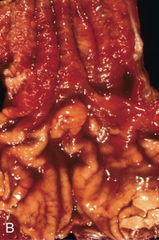
one or several tongues of salmon-pink, velvety mucosa extending upward from the gastroesophageal junction
question
Barrett esophagus
answer
metaplastic mucosa alternates with residual smooth, pale squamous mucosa of the normal esophagus and interfaces with light-brown columnar (gastric) mucosa distally
question
Barrett esophagus
answer
Three types of columnar metaplasia have been discerned: fundic, cardia, and specialized intestinal metaplasia - presence of goblet cells.
question
Barrett esophagus
answer
defined only as the presence of intestinal metaplasia
question
Barrett esophagus
answer
increased epithelial proliferation, often with atypical mitoses, nuclear hyperchromasia and
question
Barrett esophagus
answer
Dysplasia - stratification, increased nuclear-to-cytoplasmic ratio. Gland architecture is frequently abnormal and is characterized by budding, irregular shapes, and cellular crowding
question
Barrett esophagus
answer
a pre-malignant condition. - risk of transformation into adenocarcinoma correlates with the length of esophagus involved and the degree of dysplasia
question
Barrett esophagus
answer
Males 3:1 Smokers 2:1
question
Eosinophilic esophagitis
answer
chronic esophageal inflammatory disorder, allergic in nature and of unknown origin that is characterized by dense infiltration by eosinophilic granulocytes that is restricted to the esophagus
question
Eosinophilic esophagitis
answer
great majority of patients have a history of atopy (asthma, rhinitis, conjunctivitis, drug and food allergies), blood eosinophilia, increased serum IgE levels, and positive results in multiple allergy skin tests
question
Eosinophilic esophagitis
answer
endoscopic is often characteristic with transverse ridges (which can resemble the trachea) and small white plaques
question
Eosinophilic esophagitis
answer
most characteristic microscopic finding is a high number of eosinophils infiltrating the esophageal epithelium
question
Eosinophilic esophagitis
answer
Treatment strategies include the exclusion of sensitizing foods, systemic and topical corticosteroids
question
Candida esophagitis
answer
In mild cases, a few small, elevated white plaques are surrounded by a hyperemic zone in the middle or lower third of the esophagus
question
Candida esophagitis
answer
In severe cases, confluent pseudomembranes lie on a hyperemic and edematous mucosa
question
Candida esophagitis
answer
pseudomembrane contains fungal mycelia, necrotic debris and fibrin
question
Herpes esophagitis
answer
viral infection of the esophagus caused by Herpes simplex virus
question
Herpes esophagitis
answer
development of small vesicles that subsequently rupture to form discrete superficial ulcers on the mucosa
question
Herpes esophagitis
answer
Squamous epithelial cells in herpetic vesicles exhibit typical nuclear herpetic inclusions and occasional multinucleation
question
Cytomegalovirus esophagitis
answer
Characteristic cytomegalovirus inclusion bodies are seen in endothelial cells and granulation tissue fibroblasts
question
Chemical esophagitis
answer
results from ingestion of corrosive agents
question
Chemical esophagitis
answer
reflects accidental poisoning in children, attempted suicide in adults or contact with medication ("pill esophagitis")
question
Chemical esophagitis
answer
Ingestion of strong alkaline agents (e.g., lye) or strong acids (e.g., sulfuric or hydrochloric acid), both of which are used in various cleaning solutions, can produce
question
Iatrogenic esophagitis
answer
External irradiation for treatment of thoracic cancers
question
Iatrogenic esophagitis
answer
Nasogastric tubes may cause pressure ulcers when they are in place for prolonged periods, although acid reflux also plays a role in these cases.
question
ESOPHAGEAL VARICES
answer
dilated veins immediately beneath the mucosa that are prone to rupture and hemorrhage
question
ESOPHAGEAL VARICES
answer
arise in the lower third of the esophagus - virtually always in patients with cirrhosis and portal hypertension
question
ESOPHAGEAL VARICES
answer
response to portal hypertension is the development of a collateral circulation diverting the obstructed portal blood flow to the caval veins - portocaval collaterals
question
ESOPHAGEAL VARICES
answer
most important portosystemic anastomoses are the gastroesophageal collaterals
question
ESOPHAGEAL VARICES
answer
native veins laying within the wall of the esophagus and draining into the azygos vein.
question
ESOPHAGEAL VARICES
answer
appear as tortuous dilated veins lying primarily within the submucosa of the distal esophagus and proximal stomach
question
ESOPHAGEAL VARICES
answer
If exceed 0.5 cm in diameter, they tend to rupture, leading to life-threatening massive upper gastrointestinal hemorrhage.
question
MALLORY-WEISS SYNDROME
answer
bleeding from tears in mucosa at gastroesophageal junction - usually caused by severe retching, coughing, or vomiting
question
MALLORY-WEISS SYNDROME
answer
most patients present with a single tear involving mucosa and submucosa but not the muscular layer
question
MALLORY-WEISS SYNDROME
answer
On average, the tear is 2-3 cm in length and a few millimeters in width
question
Most common benign tumor
answer
leiomyomas
question
Squamous papillomas
answer
sessile lesions with a central core of connective tissue and a hyperplastic papilliform squamous mucosa.
question
SCC
answer
- thoracic esophagus - AA males - Smoking/Alcohol - in US - over 45, males 4:1
question
Adenocarcinoma
answer
- Caucasian males - linked to GERD/Barrett esophagus
question
SCC
answer
middle and upper thirds of the esophagus
question
SCC
answer
tumors may be polypoid exophytic masses that protrude into the lumen leading to obstruction or cancerous ulcerations that infiltrate deeply within the esophageal wall and gradually narrow the lumen by circumferential compression
question
SCC
answer
neoplastic squamous cells range from well- differentiated forms, characterized by presence of epithelial "pearls," to poorly differentiated forms in which the evidence of squamous differentiation is minimal or absent
question
SCC
answer
rich lymphatic drainage of the esophagus provides a route for most metastases
question
SCC
answer
tumors of the upper third metastasize to cervical, internal jugular and supraclavicular nodes
question
SCC
answer
Metastases to liver and lung are common
question
SCC
answer
Cancer of the middle third spreads to paratracheal and hilar lymph nodes and to nodes in the aortic, cardiac and paraesophageal regions
question
SCC
answer
Because the lower third of the esophagus is fed by the left gastric artery, lower esophageal tumors spread via accompanying lymphatics to retroperitoneal, celiac and left gastric nodes
question
SCC
answer
most common presenting complaint is dysphagia, but by the time a patient complains of dysphagia, most tumors are unresectable
question
SCC
answer
cachectic owing to difficulty in swallowing
question
SCC
answer
Odynophagia occurs in half of patients, and persistent pain suggests mediastinal extension of the tumor or involvement of spinal nerves
question
SCC
answer
Compression of the recurrent laryngeal nerve causes hoarseness, and tracheoesophageal fistula is manifested clinically by a chronic cough
question
ADENOCARCINOMA
answer
arises in a background of Barrett esophagus and long-standing GERD
question
ADENOCARCINOMA
answer
Caucasian men 7:1
question
ADENOCARCINOMA
answer
distal third of the esophagus and may invade the adjacent gastric cardia
question
ADENOCARCINOMA
answer
Initially appearing as flat or raised patches in otherwise intact mucosa
question
ADENOCARCINOMA
answer
Chromosomal abnormalities and mutation or overexpression of p53 are present at early stages
question
ADENOCARCINOMA
answer
amplification of c-ERB-B2, cyclin D1, and cyclin E genes; mutation of the retinoblastoma tumor suppressor gene; and allelic loss of the cyclin-dependent kinase inhibitor p16/INK4a. In other instances p16/INK4a is epigenetically silenced by hypermethylation


