Structural Principles of Osteopathic Medicine, Exam 2 – Flashcards
Unlock all answers in this set
Unlock answersquestion
Innervation of face and scalp
answer
Somatic sensory (skin, conjunctiva of eye, parotid gland used in salivating) Somatic (Branchial) motor (muscles of facial expression) Visceral motor (autonomics) ?Parasympathetics to parotid gland (source: glossopharyngeal nerve) ?Sympathetics to iris, eyelids, blood vessels, arrector pilae mm., sweat glands (source: postganglionic axons from superior cervical ganglion following arteries to targets)
question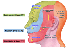
Somatic sensory innervation of the face

answer
Trigeminal nerve (CN V) has 3 major divisions, named for the region they are going to ?Superior most region - ophthalmic division ?Maxillary division exits near maxilla ?Mandibular division rides the course of the lower jaw (mandible) ?Terminal branch is the mental nerve There are also branches from cervical plexus and from dorsal rami of cervical spinal nerves
question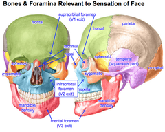
Bones & Foramina Relevant to Sensation of Face

answer
Sphenoid is bone in the back of the orbit ?From lateral view, has winging out aspect Lacrimal and nasal bones are where your tear ducts are, as well as bridge of nose Zygomatic is prominent cheek bone Maxilla and mandible are also important Supraorbital foramen is superior to the orbit Infraorbital is inferior to the orbit Mental foramen is in the mandible
question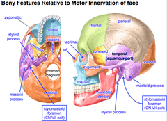
Bony Features Relative to Motor Innervation of Face

answer
Temporal bone ?Styloid process is a long process ?Mastoid process is bulbous and round ?b/w them, is the stylomastoid foramen ?CN VII motor exits here then branches while passing through parotid gland, arriving at muscles of facial expression ?DOES NOT INNERVATE PAROTID GLAND ?CN VII = facial nerve
question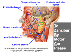
Facial Nerve

answer
To Zanzibar By Motor Car Please (acronym for remembering branches of facial nerve) To = temporal branches Zanzibar = zygomatic branch By = buccal branch Motor = mandibular branch Car = cervical branch Please = posterior auricular branch
question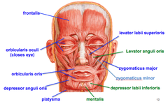
Muscles of Facial Expression

answer
Frontalis is right on frontal bone Orbicularis oculi surround the eye, allows you to squint Orbicularis oris lets you purse your lips, whistle, close mouth Depressor anguli oris comes off the corners of your mouth so you can pull it down in one direction Platysma is muscle in neck that flexes in anger/stress (aka fearsome muscle) Levator labii superioris rides along nose, if you contract it you lift lip up Zyogomaticus major and minor run together ?Work together ?Come from superior corner of mouth ?Help you smile Levator anguli oris elevates angle of mouth Depressor labii inferioris drags corners of mouth down Mentalis helps you pout your mouth
question
Scalp
answer
Frontal belly and occipital belly are connected by aponeurosis ?When you act on one, it acts on the other Going from superficial to deep ? SCALP ?Skin ?Connective tissue ?Aponeurosis ?Loose connective tissue ?Pericranium (outer periosteum)
question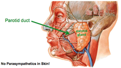
Parotid Gland

answer
Facial nerve runs through but does NOT control the parotid gland It gets parasympathetic innervation from the glossopharyngeal nerve (CN IX) ?Will be postsynaptic neurons (ganglion buried in bones of skull) Saliva travels through parotid duct ?Pierces the buccinator muscle, past the masseter
question
Development of facial muscles
answer
Take about 2 years to develop This is why infants drool ? do not yet have control of facial muscles
question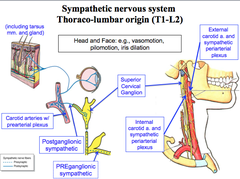
Sympathetics of Face and Scalp

answer
Sympathetics for head and neck go to blood vessels, hair, skin ?Can dilate iris so it can open up and allow more light in In skin, found in body wall (true for entire body) Pre-synaptic fibers come from T1-L2 ?White not quite, gray all the way ?Enters ganglia via the white ramus, synapses at most appropriate ganglion and goes to source via gray rami Post-synaptic sympathetics jump onto carotid arteries to get to the places that they need to go
question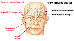
Superficial Arteries to Face and Scalp

answer
From external carotid: ?Superficial temporal artery (from maxillary artery) ?Transverse facial (zygomatic arch of cheek bones) ?Facial artery (branches at jaw, does not take totally direct path up towards nose) From internal carotid: ?Supratrochlear artery ?Supraorbital artery comes out of the supraorbital foramen
question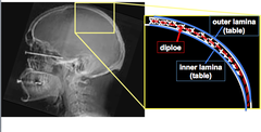
Skull

answer
Skull is spongy bone, just like with long bones Has both an outer lamina and inner lamina, w/ spongy layer in b/w called the diploe The periosteum is over the inner and outer laminae and is continuous at sutures, foramina and canals ?Lots of innervation in periosteum ? important b/c you will feel pain if its damaged
question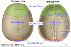
Calvaria

answer
Suture b/w frontal bone and parietal cones is the coronal suture b/w parietal bones is the sagittal suture b/w parietal bones and the occipital bone is the lambdoid suture
question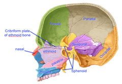
Cranial Cavity

answer
Image is approximately just left of the midline b/c skull is facing to left and you can see what is basically nasal septum of ethmoid bone ?If you were in midline, you would have busted through nasal septum Can see that part of nasal septum is created by palatine muscle, which also contributes to hard palate Can see sphenoid sinus as well Cribriform plate of ethmoid bone - small portion of the ethmoid w/ small punctures in it, where nerve endings of olfactory nerve go through, allowing smell
question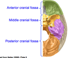
Cranial Fossae

answer
As you move from anterior to posterior, it is like walking down steps ? gets deeper and deeper Covered by dura mater
question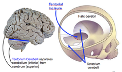
Dural Folds

answer
Create little divisions in/around parts of the brain Tentorium cerebelli separates cerebellum (inferior) from cerebrum (superior) ?Tentorial incisure is a shelf on which the cerebrum sits and the cerebellum rests underneath Falx cerebri splits cerebrum in half
question
Meningeal Arteries
answer
Meningeal arteries are embedded w/in endocranium Middle meningeal artery lies deep to pterion ?supplies most of cranial dura and bones of calvaria
question
Pterion

answer
Sutural intersection formed by: ?Frontal bone ?Parietal bone ?Squamous temporal ?Greater wing of sphenoid Overlies anterior branches of middle meningeal artery This area is very thin ? weak zone ?If you break these bones, you may damage blood vessels and create an epidural hematoma
question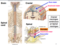
Meninges of Brain

answer
Cranial meninges are continuous w/ spinal meninges Basically the same, but in the brain the dura folds upon itself
question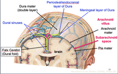
Cranial Meninges

answer
Dura folds upon itself ? 2 layers of dura ?more superficial is against the periosteum layer of bone (periosteal layer of dura) ?deeper layer is up against the next meningeal layer (meningeal layer of dura) arachnoid mater overlies CSF pia mater is right up against nerve tissue
question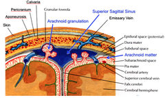
CSF Recycling

answer
CSF punches through the meningeal layer of the dura at the meningeal sinuses Arachnoid matter punches through dura to make dural sinuses ?CSF can be recycled through this zone via arachnoid villi ?Venous drainage occurs here too Arachnoid villi can leave granulation scars on inner surface of skull
question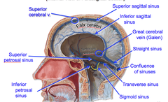
Dural Folds and Venous Sinuses

answer
Superior sagittal sinus drains blood from superior cerebral veins Inferior sagittal sinus combines w/ the great cerebral vein to form straight sinus Straight sinus merges with superior sagittal sinus to form the confluence of sinuses Then moves out along the transverse sinus, picking up flow from superior petrosal sinus on the way Ultimately exits via jugular vein Cavernous sinus is basically giant cave in skull w/ convoluted blood vessels (mainly venous) • Home of pituitary gland
question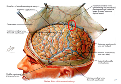
Dural Sinuses - Venous Drainage

answer
Cerebral veins punch into superior sagittal sinus Can have shearing force which busts superior cerebral veins and causes subdural hematoma ?Happens before vein punctures arachnoid mater
question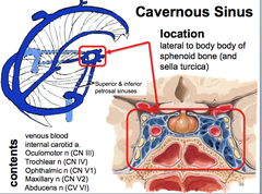
Cavernous Sinus

answer
Mostly convoluted venous blood Cooler blood here helps to cool down arterial blood as it enters this area to supply the brain Have many different nerves here ?Oculomotor nerve moves the eye ?Trochlear nerve moves the eye ?Ophthalmic nerve provides sensory info from nerve ?Maxillary nerve sensory info from eye ?Abducent nerve abducts eye
question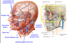
Superficial Veins of the Face and Scalp

answer
Veins of the face drain back towards the cavernous sinus ?Potential source of infection
question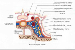
Cavernous Sinus Syndrome

answer
Impingement of nerves in cavernous sinus Complains of numbing of eye Has problems moving eye
question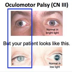
Oculomotor Palsy (CN III)

answer
Impingement of nerves in cavernous sinus Complains of numbing of eye Has problems moving eye
question
Horner's Syndrome
answer
Sympathetics knocked out to the head Group of symptoms that make syndrome ?Tosis (drooping of eyelid) ?Myosis (constricted pupil) ?Anhydrosis (lack of sweating)
question
Facial Development Timeline
answer
Origin: 4th week, completed at 8 weeks
question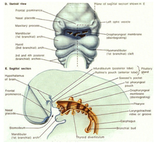
The face arises from 5 mesodermal primordia

answer
Two maxillary prominences from branchial arch I Two mandibular prominences from branchial arch I One frontonasal prominence from the forehead In addition, paired nasal placodes (ectodermal) form in frontonasal prominence and each nasal placode becomes surrounded by medial and lateral nasal prominence of mesodermal origin
question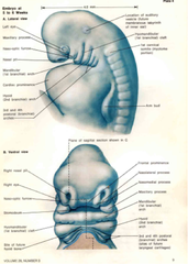
Facial Growth

answer
Maxillary prominences approach each other and the nasal prominences ventromedially Medial nasal prominences fuse in the midline and also w/ the maxillary prominences ?Lateral nasal prominence and maxillary prominence on each side remain separated by a cleft called masolacrimal groove ?Ectoderm in this groove forms the nasolacrimal duct and lacrimal sac ?Common anomaly occurs when the ectoderm fails to hollow out so that the nasolacrimal duct remains atretic Two mandibular prominences fuse medially
question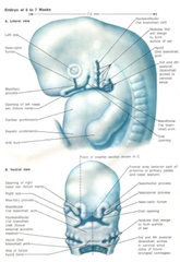
Derivatives of Facial Primordia

answer
During the 6th-7th week, differentiation occurs as follows: ?Mandibular prominences form the lower jaw, lower lip, lower part of face ?Frontonasal prominence forms forehead and dorsum ; apex of nose ?Lateral nasal prominences - form sides of the nose ?Medial nasal prominences - fuse to form intermaxillary segment of the upper jaw (philtrum, gingiva, primary palate and middle of upper jaw) and the nasal septum ?Maxillary prominences form lateral parts of upper lip ; jaw and secondary palates
question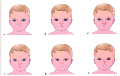
Craniofacial Clefts

answer
Results when mandibular and/or maxillary prominences fail to merge or when the maxillary prominence fails to fuse w/ lateral and medial nasal prominences In latter case, an oblique facial cleft results which runs from the eye to the mouth on the same side When mandibular prominences fail to fuse completely a median cleft of the lower jaw occurs and when the mandibular and maxillary prominences fail to merge correctly on the same side, a lateral facial cleft results ?Latter causes large mouth extending back almost to ear ? most common of facial clefts
question
Bifid Nose
answer
Results form failure of medial nasal prominences to merge in the midline As a result, nose remains split
question
Palate Development Timeline
answer
Forms during 5th-12th week
question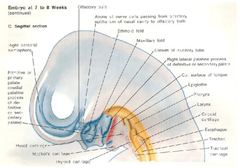
Development of the Palate

answer
Palate is derived from 2 parts: primary palate and secondary palate Primary Palate (median palatine process) ?Arises as a mesodermal thickening of the innermost (premaxillary) part of the intermaxillary segment of the upper jaw during the 5th week ?Located b/w maxillary prominences of the upper jaw Secondary Palate (lateral palatine process) ?Arises as mesodermal projections from inner surface of each maxillary prominence ?Initially they project down each side of tongue, but as jaw develops the tongue moves inferiorly allowing the lateral palatine processes to grow together and fuse above the tongue ?Fusion in the midline begins during the 9th week and involves the secondary palates fusing with: ?Primary palate ?Nasal septum (derived from medial nasal prominences) ?Fusion is complete by end of 12th week ?Proceeds in an anterior-posterior direction, leaving palatine raphe as a fusion line
question
Bone formation in the palate
answer
Primary palate - membranous bone formation gives rise to the premaxillary part of the upper jaw (for incisor teeth) Secondary palate - membranous bone gives rise to hard palate ?No bone forms in the secondary palate posterior to the nasal septum ? becomes soft palate and uvula
question
Cleft Lip
answer
May be unilateral or bilateral ?Unilateral - maxillary prominence on one side fails to merge w/ medial nasal prominence on the same side, resulting in persistent labial groove ?Epithelium in groove becomes stretched, and it and the underlying mesoderm break down, and the lip divides into a medial and lateral part ?Bilateral - medial nasal prominences fuse but maxillary prominences fail to meet and fuse with the fused medial nasal prominences ?Epithelium in both grooves now stretches and it and the underlying mesoderm breaks down causing bilateral cleft lip 1/800 - 1/900 births ?more common in males
question
Cleft Palate
answer
Results from the failure of the lateral palatine processes to meet and fuse either with a) each other, b) nasal septum) or c) the median palatine process May involve only the uvula or the soft and hard palate as well Cleft lip may or may not be associated with cleft palate Cleft of Primary Palate - the lateral palatine processes fail to fuse with the median palatine process Clefts of Primary ; Secondary Palates - lateral palatine processes fail to fuse with each other, the nasal septum and the median palatine process Clefts of the Secondary Palate - lateral palatine processes fail to fuse with each other and with the nasal septum 1/2500 births ?more common in females
question
Nasal Cavity Development Timeline
answer
Origin: 4th week
question
Development of the Nasal Cavities
answer
Begins as two ectodermal swellings (nasal placodes) induced by the forebrain Nasal placodes become pits as the surrounding mesodermal medial and lateral nasal prominences grow up around them Pits are constricted off to form sacs which communicate with the surface via the nasal pares Initially the sacs are separate, but fuse together to form one cavity separated from the oral cavity by the oronasal membrane ?Oronasal membrane ruptures at 6 weeks and the openings become the choanae The oral pharynx and nasal pharynx are secondarily separated again by the primary and secondary palates (except at choanae) and the nasal cavity is separated into left and right nasal cavities by the nasal septum Olfactory region ?Ectodermal lining from ingrown nasal placodes form specialized olfactory cells (neuro-sensory, ciliated) which send fibers (i.e. nerve fibers that comprise the olfactory cranial ns) to synapse in the olfactory bulb
question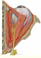
Contents of the Orbit

answer
Eyeball Nerves Ocular muscles Fascia Vessels Lacrimal gland and sac ?Consider for dry eye Fat (cushions eyeball)
question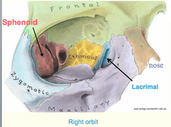
Bones of the Orbit

answer
?Frontal ?Sphenoid ?Broken up into divisions - Lesser wing and greater wing (must know and identify) ?Zygomatic ?Maxillary ?Lacrimal ?Ethmoid Clinical relevance: if there's a tumor, you need to think about what bones are present and which nerves, arteries and glands might be in the area and would be affected
question
Walls of Orbit

answer
Superior - frontal bone Lateral Wall - sphenoid & zygomatic bones Inferior wall - maxillary and zygomatic bones Medial wall - lacrimal & ethmoid bones
question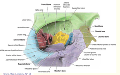
Frontal Bone in the Orbit

answer
Supraorbital notch ?Superior orbital nerves and vessels go through this notch ?Supraorbital vessels and nerve (V1 branch of ophthalmic) ?If patient was hit in the head with a ball, have loss of sensation here (forehead) there might be damage to V1 Anterior ethmoidal foramina ?Anterior ethmoidal vessels and nerve (V1 branch of ophthalmic) ?Innervate ethmoid sinuses ?Think pain and sinus pressure Posterior ethmoidal foramina ?Posterior ethmoidal vessels & nerve (V1 branch of ophthalmic) ?Innervate ethmoid sinuses ?Think pain and sinus pressure
question
Maxillary Bone in the Orbit
answer
Infraorbital groove ?Infraorbital nerve (V2 branch of maxillary nerve) goes through this groove, through canal and goes through foramen ?Major sensory nerve to upper teeth, upper lip, lower part of eye ?Infraorbital artery
question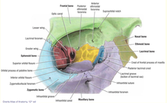
Lacrimal bone in the orbit

answer
Lacrimal groove ?Lacrimal sac (reservoir for tears) sits here ?This is what you would massage in a newborn if they have blocked tear ducts ?May be a tumor in lacrimal groove, buildup of bacteria, etc.
question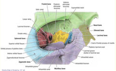
Sphenoid Bone in the Orbit

answer
Less wing and greater wing separated by infraorbital fissure and superior orbital fissure This is where the trigeminal, trochlear, abducens and oculomotor nerves are located Optic canal ?Optic nerve (CNII) ?Ophthalmic artery Inferior orbital fissure ?Trigeminal nerve (V2, maxillary division) ?Infraorbital artery Superior orbital fissure ?Trigeminal CN (V1) (ophthalmic division) ?Oculomotor nerve (CN III) ?Trochlear nerve (CN IV) ?Abducens nerve (VI) (superior and inferior division) ?Ophthalmic vein
question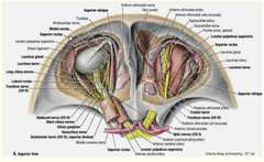
Nerves in the Orbital Cavity

answer
In superior view of orbit, the first things you will see are muscles of eye involved in eye movement ?Superior oblique muscle also visualized Major nerve to see in superior portion is the frontal nerve ?Branches to supraorbital nerve and supratrochlear nerve Optic nerve forms the optic chiasm ?NEED TO BE ABLE TO IDENTIFY Can also see the nasociliary branch of the trigeminal nerve ?Gives off the long ciliary nerves (sensory) ?Short ciliary nerves comes off of the ciliary ganglion (which hangs onto nasociliary) Ciliary ganglion carrier parasympathetic division of CN III involved in constriction of pupil ?Transmits up into ciliary muscle involved in pupillary reflex
question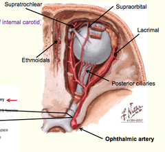
Arteries of the Orbital Cavity

answer
Major artery is the ophthalmic artery ?The internal carotid gives off ophthalmic artery ?Branch of ophthalmic artery goes through dural sheath and gives off central retinal artery Central retinal artery is major arterial supply of cells that line eye and are responsible for transmitting light signals ?If you have massive compression on optic nerve at a point where the retinal artery has entered, you will impinge blood supply as well, could be problem of visual system Other branches of ophthalmic artery are: ?Supraorbital ?Supratrochlear ?Lacrimal ?Ethmoidals ?Posterior ciliaries
question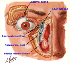
Lacrimal Apparatus

answer
Lacrimal gland ?Sensory from trigeminal nerve ?Secretes water, mineral salts, antibodies and bactericidal enzymes ?Want this gland to function to prevent infection You have a flow source into lacrimal canaliculi ?Gets blocked when you have a cold Drains to lacrimal sac ?Reservoir ?You get the sniffles because it drains to nasolacrimal duct, and then your nose drips
question
Eyelid Muscles

answer
Orbicularis oculi (3 segments, all circular) ?Palpebral ?Light closure of eyelid ?Involved in unconscious blinking ?Orbital ?Tight closure of eyelid ?Conscious squinting ?Lacrimal ?Draws eyelids medially, squeezes lacrimal gland Levator palpebral ?Lifts eyelid ?Runs in a vertical direction ?Innervation by oculomotor cranial nerve ?Important in patient w/ eyelid drooping, can quickly identify a nerve that is not working properly
question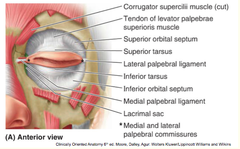
Fascia of the Eyelid

answer
Orbital septum ?Skeleton of eye ?Where muscles attach to move eyelid ?Superior and inferior portion of fascia Palpebral Ligaments ?Medial and lateral ligaments ?Important to keep eyeball in place
question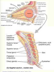
Glands in the Eyelid

answer
Tarsal glands ?Embedded in tarsal plates ?Lubricate edges of eyelids Ciliary glands ?Found in margin of eyelid ?Modified sweat glands Both tarsal and ciliary ?Secrete lipids to seal in fluid of eye ?Implications in dry eye ?Mascara can clog these glands
question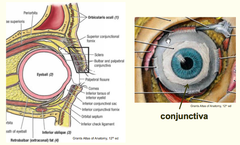
Conjuctiva

answer
Walls of the front of eyeball Invests into cornea Lines anterior portion of eye and interior of eyelid Prevents bacteria from getting into eye so things don't get into brain
question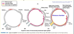
Layers of eyeball

answer
Cornea ?Transparent, important for visualizing ?Fibrous and avascular ?This has 5 layers made of collagen and epithelial tissue Sclera ?Highly vascularized ?Is white of eye ?Fibrous and thick Choroid layer ?Highly vascularized ?Helps get arterial supply from ophthalmic artery and feeds to retinal layer Aqueous humor is in anterior region of eye, b/w iris and cornea ?Includes pupil Posterior chamber (vitrous humor) is posterior to iris of eye Canal of Schlemm ?Important in glaucoma ?Older patients w/ diabetes can et this canal of schlemm clogged, which causes pain and intraocular pressure/pain
question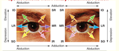
Extraocular Eye Muscles

answer
Innervations ?Lateral rectus 6 ?Superior oblique 4 ?All others innervation by CN 3 Recti Orbital muscles ?insert on sclera ?Superior rectus ?superior part of eyeball, ?CN III ?Elevates, adducts and rotates eyeball medially ?Inferior rectus ?inferior part of eyeball ?CN III ?Depresses, adducts, and rotates eyeball medially ?Medial rectus ?medial part of eye ball (note the nose) ?CN III ?adducts eyeball ?Lateral rectus ?lateral part of eyeball, ?CN VI ?Abducts eyeball ?These all have the same origin: the Common Tendonous Ring Obliques Orbital muscles ?Superior Oblique ?Origin = sphenoid bone ?CN IV ?Abduct, depresses and medially rotates eyeball ?Inferior oblique ?Origin = maxillary bone ?CN III ?Abducts, elevates and laterally rotates eyeball These movements are from primary neutral position which is the pupil forward and center. The muscles work in yolked pairs Note the difference between anatomical and clinical ?Clinical is the H test
question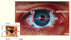
H test

answer
Two muscles depress eye, two muscles that elevate To differentiate, you must take to extreme position. Follow your finger to extreme position. Then look up and down Example: Right eye ?Follow to nose and down: testing superior oblique (Could also be testing CN IV).. Because SO4 ?Follow to lateral and then down... testing inferior rectus
question
Types of Connective Tissue
answer
Embryonic (allow for a lot of flow/movement b/c circulatory system is not fully developed) ?Mesenchyme ?mucous Adult ?Loose (Areolar) connective tissue ?Dense connective tissue ?Dense regular connective tissue ?Dense irregular connective tissue ?Reticular connective tissue ?Elastic connective tissue Special ?Adipose ?Hematopoietic tissue ?Lymphatic tissue (most of lymphatic organs are composed of connective tissue) ?Elastic ?mucous Supporting ?Cartilage ?Bone
question
Functions of Connective Tissue
answer
Supporting tissue ?Hooks up cells and fibers and ground substance Packing material Nutrition Exert or resist force (tensile strength) ?Everything is interconnected Storage Defense ?when there is infection, it usually winds up in the connective tissue
question
General Features of Connective Tissue
answer
Mostly extracellular matrix ?Fibers ?Ground substance (non-fibrous portion) Cells ?Fibroblasts ?Macrophages ?Mast cells ?Leukocytes ?Plasma cells ?Adipocytes ?Mesenchymal
question
Fibers of Connective Tissue
answer
Collagen (main fibrous component) ?Most abundant protein of the human body ?7 distinct collagen ?-chains, each about 1000 AAs long ? triple strand helical structure ?AAs: Glycine (every 3rd AA), proline, hydroxyproline, hydroxylysine Reticular Elastic
question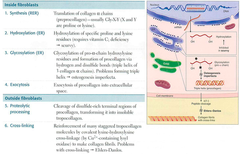
Collagen Synthesis

answer
When you put collagen together, you want to make sure it forms properly ?Don't want it to form within a cell, b/c the cell will become fibrotic and die Make pre—procollagen in the cell (w/ signaling and registration peptides) ?Becomes procollagen once the signaling peptide is cleaved off ?Registration peptides prevents folding Tropocollagen is assembled in the extracellular space, once the protein has been transported out of the cell ? fibrils can form
question
Types of Collagen
answer
Type I - bone, tendon ?Most connective strength Type II - hyaline and elastic cartilage Type III - reticular fibers Type IV - basal lamina Type V - amnion and chorion in the fetus Type VII - anchors basal lamina to stroma ?Kind of rare, but it is important in terms of interconnectivity of basement membrane w/ parenchyma ?Mutation here can mean that epidermis will not adhere to underlying dermis, leading to easy bruising, tearing ? usually don't live past 20-30's
question
Collagen Disorders
answer
Type I Collagen ?Osteogenesis imperfecta ?Progressive systemic sclerosis ?Scurvy Type II ?Hypochondrogenesis ? malformation of cartliages in bony tissues ?Short in stature, malformed facial features Type III ?Ehler-Danlos (type IV) ?Reticular fibers support things like blood vessels, so individuals w/ type IV are subject to bruising b/c blood vessels are not supported the way they should be Type IV ?Alport syndrome ? syndrome that affects basement membrane ?Organs where the basement membrane is important are usually affected, i.e. kidneys, eyes (blindness b/c passage of light is blocked by thickened basement membrane), ears Type V ?Ehler-Danlos (type I) ?While Type V is usually fetal tissue, but when it is present in adult tissue it makes the tissue less rigid
question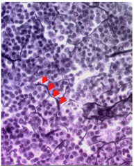
Reticular Fibers

answer
Type III collagen Affinity for silver stains Provides supporting meshwork in cellular tissues (i.e. liver, bone marrow, lymphoid organs, spleen) ?Cellular organs that don't have space for big patches of connective tissue use reticular fibers as scaffolding system
question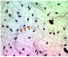
Elastic Fibers

answer
Do not contain collagen Comprised of protein elastin, microfibril-associated glycoproteins, and fibrillin Uncommon amino acids (desmosine and isodesmosine) cross link mature elastic fibers ?Important for preventing elastic fibers from just stretching until they break ?Create strength fir elastic fiber itself In blood vessels ? want blood vessels to be able to stretch, but not stretch so much that they burst
question
Marfan Syndrome
answer
Fibrillin disease ? elastic tissue disease w/o cross-linkers to limit stress, does not give fibers any resistance ?sometimes blood vessels stretch too thin, and are in danger of rupturing medical concerns of manifest around high school and college age ? tend to push bodies more then, potentially causing ruptured vessel
question
Ground Substance of Connective Tissue
answer
Gel-like substance Glycosaminoglycans (GAGs) ?Hydrophilic ? hold water ?Each polysaccharide is long, unbranched chain ?Repeating disaccharide units ?7 different GAGs Proteoglycans ?A little different than a typical glycoprotein (larger, less branched) Structural proteins ?Fibronectin and Laminin help to link cell to ground substance ?Chondronectin ?Tenascin ?Vitronectin ?Entactin
question
Mucopolysaccharidosis
answer
Lysosomal disease related to ground substance ?Does not properly break down glycosaminoglycans, causing buildup and damage to lysosomes/cells
question
Basal lamina and reticular lamina form the basement membrane
answer
Sheet like-arrangement of extracellular matrix Interface b/w connective tissue and parenchymal cells (e.g. epithelia, muscle, nerve) Comprised of type IV collagen (mostly), type VII collagen (to connect to underlying tissue), proteoglycans, fibronectin, laminin, entactin
question
Fibroblasts

answer
Spindle-shaped, w/ elliptical nucleus Function: produce components of extracellular matrix
question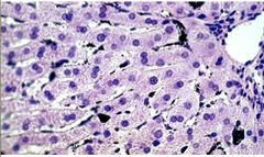
Macrophages

answer
Irregular shaped nucleus Phagocytic vesicles Abundant lysosomes Part of the immune system ? there as one to clean up cellular debris, help remodel tissues, also antigen-presenting cells
question
Special Macrophages
answer
?Kupffer cell ?Microglia ?Langerhans cell ?Dendritic cell ?Osteoclast
question
Functions of Macrophages
answer
?Ingestion of particles ?Destruction of aged erythrocytes ?Extrahepatic bile production ?Iron and Fat metabolism ?Turn over of fibers and ECM ?Antigen presenting cells ?Production of cytokines
question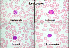
Leukocytes

answer
i.e. neutrophils, eosinophil, basophil, lymphocytes fight infection
question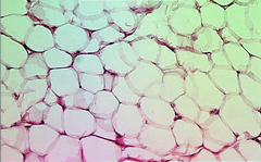
Adipose tissue

answer
Packing tissue White adipose tissue is uniocular ? one big droplet ?Looks like chicken wire
question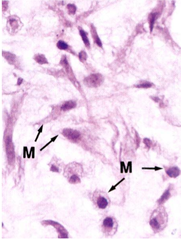
Mesenchymal cells

answer
Have totipotent/pluripotent cells that can differentiate into a variety of things ?Most will become fibroblasts
question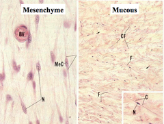
Mesenchyme and Mucous Embryonic Connective Tissue

answer
Lots of pale ECM in fetal tissues, not a lot of cells In the umbilical cord, there is wharton's jelly Lots of water for free exchange of nutrients
question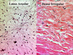
Loose Areolar vs. Dense Irregular Connective Tissue

answer
Usually a transition as you go from epithelium (loose connective) to dense connective tissue Loose tissue: more cells, less fibers Dense: more fibers, few nuclei
question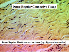
Dense regular connective tissue

answer
Collagens and fibers all tend to line up Only wants to be stretched in one direction
question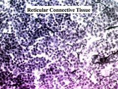
Reticular Connective Tissue

answer
Need to specifically stain for reticular fibers ? network of dark irregular lines running through very cellular tissue
question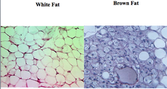
Special Connective Tissue

answer
Adipose ?White fat (single lipid droplet) ?Brown fat (multiple lipid droplets) ?Not found too often in humans ?Breaks up nuclear transport chain ? instead of generating ATP, energy is released as heat
question
Cartilage

answer
Tissue that absorbs shock, pressure ?Found covering bones that come together at a joint, symphyses, etc. ?Arranged in a way to accept force and dissipate it Chondroblast (developing cell) - produce matrix Chondrocyte (mature) - surrounded by matrix ?Chondroblasts are considered mature when they are fully surrounded Lacunae - matrix cavity Isogenous groups - all cells from mitotic division of same cell ?Cartilage is set up in a way (b/c it is fluid) that it can allow cells to multiply and separate from each other ?Will see mitotic division on slide Territorial matrix vs. ground substance ?Territorial matrix - darker, denser substance (freshly secreted from cell)
question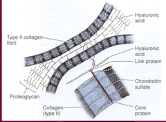
Cartilage: Extracellular Matrix

answer
Fibrils (collagen type II and I, elastin) ?Cartilage defined by presence of type II (only found in cartilage, nothing else) Ground Substance: ?Hyaluronic acid ?Lines up w/ proteoglycans w/ GAG side chains on type II collagen fibrils ?Glycosaminoglycans (chondroitin sulfate, keratin sulfate) ?Love water, love to pull water into them ?Creates a tunnel b/w collagen fibers that is very happy to receive water ? sphere of solvation SHOCK ABSORBER ?Proteoglycans ?Glycoproteins (structural links) ?Common is chondronectin
question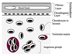
Perichondrium

answer
Actually border b/w cartilage and whatever tissue is outside of it ?Forms interface b/w cartilage and tissue supported Vascular supply for cartilage ?b/c of presence of fluid of cartilage, you have diffusion of nutrients in/out of cells ?can be a problem, b/c there is no blood supply directly into cartilage Supply of chondroprogenitor cells Outer layer - dense irregular connective tissue and fibroblasts ?Looks more regularly arranged than some, but it is NOT tendon Inner layer - chondroblasts (cells start to look fat and rounded) ?Gives rise to chondroprogenitor cells (needed to increase girth of cartilage)
question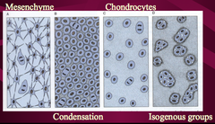
Formation of Cartilage

answer
Histogenesis - derived from mesenchyme ?Get these undifferentiated mesenchymal cells (stellate cells) which start to round up and condense ? tells cells to become chondrocytes ?Under control of TFs, growth factors, etc. ?Begin to form matrix and separate to become mature chondrocytes ?Then undergo interstitial growth ? mitotic division and growth length wise to form isogenous groups
question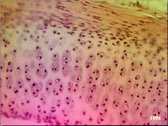
Hyaline Cartilage

answer
Most abundant type of cartilage Fibrils: collagen type II Ground substance: mostly GAGs, proteoglycans (linked by hyaluronic acid), and gylcoproteins (chondronectin) Found in: ?Fetal skeleton ?Articular surface of moveable joints (no perichondrium) ?Gets nutrition from synovial membrane ?Sternal ends of ribs ?Trachea ?Larynx ?Nose ?Epiphyseal plate
question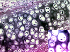
Elastic Cartilage

answer
Fibrils: collagen type II and elastin Ground substance: similar to hyaline cartilage Found in: ?External ear ?External auditory meatus ?Auditory tubes ?Epiglottis Image has dark staining, highly branched fibers ? elastic fibers
question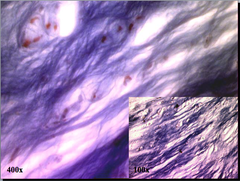
Fibrocartilage

answer
Fibers: collagen type I Ground substance: similar to hyaline cartilage No perichondrium Found in places where you bring bones together ?Intervertebral discs (anulus fibrosus) ?Insertions of tendons and ligaments into bone (make sure you can see the difference) ?Symphysis pubis Function is to take heavy levels of stress ?Fibers have to line up in direction of stress ? collagen type I is best suited for this Distinct look to it ?See cells separated out from each other, in lacunae that are not as obvious as other types of cartilage ?Look for very few cells, fibers lined up more directly, always look for an isogenous group
question
Cartilage vs. Bone
answer
Similarities ?Firm tissue that resists mechanical stress ?Cells lie in lacunae ?Mainly extracellular matrix Differences ?Bone is a hard, mineralized tissue with bony salts in matrix ?Nutrients cannot diffuse through the salt ?Presence of vasculature (b/c nutrients cannot diffuse through bone) ?Lacunae linked by canaliculi ?Collagen fibrils organized into lamellae in bone ?Appositional growth only ?Cannot have isogenous groups in bone, b/c it has no where to go
question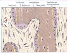
Bone Cell Types

answer
Osteoblast - secrete matrix ?Type I collagen, proteoglycans, glycoproteins Osteocyte - surrounded by matrix (mature once fully surrounded) ?Reside in lacunae ?Canaliculi are cytoplasmic processes Osteoclast - resorb matrix ?Multinucleated giant cells from bone marrow ?Come into bone when needed and create ruffled bone (invaginations of plasma membrane) that is filled w/ enzymes that will chew away at bone, releasing matrix components to be recycled ?Remodel bone
question
Extracellular Matrix of Bone
answer
Inorganic components ?Calcium and phosphorus ?Hydroxyapatite crystals [Ca10(PO4)6(OH)2] ?Non-crystalline hydroaxyapatite ?Magnesium ?Bicarbonate ?Citrate ?Sodium ?potassium Organic components ?Type I collagen ?GAGs ?Chondroitin sulfate ?Keratin sulfate ?Proteoglycans ?Glycoproteins (that promote calcification of matrix, i.e. osteonectin)
question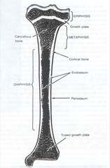
Bone: Periosteum and Endosteum

answer
Periosteum ?Outer layer = collagen fibers and fibroblasts ?Inner layer - osteoprogenitor cells Endosteum ?Single layer of osteoprogenitor cells ?Biggest source of osteoprogenitor cells ?Lines all internal cativites of bone
question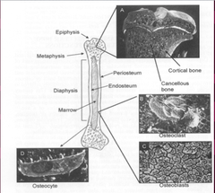
Bone Terms

answer
Metaphysis - where bones come together Epiphysis - covered w/ cartilage (no perichondrium) Diaphysis - shaft of bone
question
Primary and Secondary Tissue of Bone
answer
Primary bone tissue ?Random deposition of collagen fibers ?Lower mineral content Secondary bone tissue ?Lamellar collagen fiber arrangement ?Haversian system (osteon)
question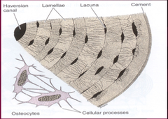
Lamellar Pattern of Bone

answer
Bone forms around blood vessels in circles (lamellae), growing as they go out further Haversian system - one unit of bone laid down circumferentially around blood vessels ?Haversian canal ?Volkman's canal - connect 2 Haversian systems Outer circumferential lamellae Inner circumferential lamellae (closest to blood system) Interstitial lamellae (closest to neighboring system)
question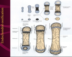
How is bone made?

answer
All flat bones are made by intermembranous ossification ?Direction mineralization of matrix secreted by osteoblasts ?Similar to histogenesis of cartilage ?Primary center of ossification: mesenchymal cells into osteoblasts All long bones are formed by endocondral ossification ?Creation of bone on cartilage scaffold ?Start w/ histogenesis of cartilage, then you get a little collar that differentiates bone out (bony collar), have primary ossification center in diaphysis, secondary ossification center in epiphysis, and growth plates at joint
question
IO Abnormalities
answer
Craniostenosis ?Crouzon Syndrome ?Cranial synostosis, exophthalmos short upper lip ?mutation in fibroblast growth factor receptor 2 ?Apert Syndrome ?Skull and midfacial abnormalities ?Syndactyly (two or more digits fused together) ?Mutation in fibroblast growth factor receptor
question
EO Abnormalities
answer
Osteopetrosis ?Increased bone mass ?Bone appears thickened in radiographs ?Abnormal osteoclast Osteogenesis imperfecta ?"brittle bone" disease ?abnormality in type I collagen ?generalized osteopenia in radiographs ?abundance of disorganized bone in histology
question
Bone Joints
answer
Synostosis - bones united by bone Synchondroses - bones joined by hyaline cartilage ?Symphysis = fibrocartilage Syndesmosis - bones connected by interosseous ligament or fibrous membrane Diarthroses - ligaments and connective tissue capsule maintains ends of bones in contact
question
Transgenic Mouse Modeling
answer
Excellent method to study complex regulatory systems Particularly important for analysis of cellular response to extracellular matrix Cellular response to external environment is based on differentiation state Knockouts are homologous recombination ?Targeted disruption of a gene ?Introduced into single-cell embryos and transgenic lines established Tissue-directed transgenes ?Promoter that has tissue-restricted activity ?More specific targeting of the knockout Possible outcomes ?No phenotype ?Redundancy of target molecule ?Mild phenotype ?Redundancy compensates ?Severe phenotype ?Prenatal lethality ?Targeted disruptions ?Tissue-specific promoters Mice containing an OI producing glycine substitution in collagen type I ?Perinatal lethal OI ?As little as 10% expression of transgene ?Mutant ECM Mice containing a large internal deletion in the collagen type I molecule ?Less severe OI ?Correlated with degree of transgene expression ?Few mutant proteins in ECM
question
Achondroplasia
answer
Impaired maturation of cartilage in the developing growth plate Affects all bones formed from cartilage Major cause of dwarfism Shortening of proximal extremities, bowing of legs, lordosis
question
Osteogenesis Imperfecta
answer
Brittle bone disease Abnormal development of type I collagen Multiple bone fractures Blue sclera Hearing loss, abnormal dentition
question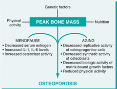
Osteoporosis

answer
Decrease in bone mass ?Bony trabeculae thinner and more widely separated Structural changes lead to increased bone fragility ?Affects femoral necks and vertebrae Hormonal factors ?Estrogen deficiency may increase bone loss and decrease bone synthesis Genetic factors: VDR molecule Mechanical factors - less weight-bearing, exercise Role of diet - need calcium in diet
question
Osteomalacia
answer
Aka rickets In children with open epiphyseal plates produces cup shaped deformity on x-ray Accumulation of unmineralized bone by defective mineralization Decreased serum calcium or phosphate Due to decreased levels of vitamin D
question
Parathyroid Hormone
answer
Osteoclast activation: increased bone resorption and calcium mobilization Increased resorption of calcium by renal tubules Increased synthesis of vitamin D
question
Hyperparathyroidism
answer
Bone resorption Replacement of bone by loose connective tissue Erosion of bone surfaces Primary: autonomous secretion Secondary: chronic renal insufficiency
question
Osteomyelitis
answer
Bone infections, can be particularly difficult to treat b/c of restricted blood supply ?May need prolonged antibiotic therapy ?May need to surgically drain infection Bacterial: mostly staphylococcus aureus, maybe e. Coli, klebsiella or proteus Salmonella: sickle cell pts H. Influenza: newborns Pseudomonas: IVDA (IV drug abusers, prone to organisms that general population may not be) Tuberculosis: Pott's disease ?Hematogenous spread ?Direct extension from joint or tissue ?Traumatic implantation from surgery or trauma
question
Acute Osteomyelitis
answer
Neutrophilic inflammation Necrosis (variable degrees) Subperiosteal abscesses Disruption of blood supply Spread to joint capsule
question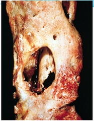
Chronic Osteomyelitis

answer
Sequela of acute infection Sequestrum: residual necrotic bone Involucrum: rim of reactive bone Brodies abscess: abscess surrounded by sclerotic bone Bacteria may be present in sequestered area
question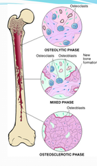
Paget's Disease

answer
Most patients ;55y/o More common in England, Australia, N. Europe Usually affects lumbar sacral area spine, pelvis, femur, skull Research shows probably viral etiology May develop secondary osteosarcomas Usually asymptomatic, but some symptoms are: ?Serum alkaline phosphatase often elevated from osteoclastic activity ?Hypervascular bone lesions: warm skin, increased cardiac output ?Enlargement of head: headaches, visual disturbances, deafness ?Transverse fractures of long bones Osteolytic phase: marrow replaced by connective tissue w/ osteoclasts Mixed phase: bone resorption and bone formation Osteosclerotic phase: irregular bone deposition causing a mosaic pattern
question
Osteomas
answer
40-50 y/o males ; females exophytic lesion of dense mature bone on flat bones of skull and face ?may protrude into sinuses Associated with Gardner's syndrome May cause cosmetic problems Not malignant
question
Osteiod Osteomas
answer
10-30 y/o Male > female Femur, tibia in metaphysis Painful ?Relieved by aspirin Central radiolucent nidus High levels of prostaglandins
question
Osteoblastoma
answer
Similar to osteoid osteoma Larger central nidus Spine, large bones of legs Often painless May be difficult to distinguish from osteosarcoma
question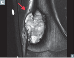
Osteosarcoma

answer
Most common primary bone malignancy (except hematopoietic lesions) Male:female ratio is 3:2 Usually 10-25 y/o w/ second peak after 40 years Usually metaphysis, lower femur and upper tibia Malignant cells form osteoid Extend from marrow to cortex to soft tissue to epiphysis to joint Codman's triangle: elevation of periosteum May have satellite nodules May destroy pre-existing bone or grow around it Usually metastasize via blood ?Lung most common site ?May spread to other bones ?Regional lymph nodes almost never involved
question
Osteochondroma

answer
Aka exostoses Approximately 10 y/o Metaphysis, lower femur, upper tibia, humerus Grows in opposite direction of joint Cap of cartilage w/ bone underneath Most asymptomatic May spontaneously regress Rare malignant transformation if single ?Multiple may be associated w/ Gardner's syndrome ?If multiple, more likely to undergo malignant transformation
question
Overview of Integument
answer
Epidermis ?Thick and thin Dermis ?Dermal papillae Epidermal derivatives ?Hair, glands and nails Hypodermis (aka fascia) ? what holds integument to rest of body
question
Functions of Integument
answer
?Prevent dessication (process of extreme drying) ?Communication ?Protection (injury and UV) ?Thermoregulation, metabolism and excretion ?Expansion ?Dermatoglyphics (fingerprinting)
question
Overview of Epidermis
answer
Stratified squamous keratinized epithelium Cell types ?Keratinocytes - cells of epithelium of integument (ectodermal derivative) ?Melanocytes - has to do w/ producing melanin, skin tone (neural crest derivative) ?Langerhan's cells - antigen-presenting cells ?Merkel's cells - free nerve endings Thick vs. thin skin ?Depends on location of skin in question ?Thin skin can support hair follicles, whereas thick skin cannot b/c of thick layer of stratus corneum
question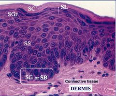
Epidermal Layers

answer
From connective tissue ? surface Stratum basale (germinativum) - layer that renews and gives rise to stem cells that replace the skin ?Replace entire layer of skin every 15-30 days Stratum spinosum - looks like there are spiny processes on edges of cell ?Malpighian layer - where all mitotic activity is ? b/w basale and spinosum Stratum granulosum - dark, granular staining material, keratohyalin granules Stratum lucidum (in very thick skin) - clear, cells begin to flatten out, losing organelles, beginning to die, packed keratin tonofilaments Stratum corneum - very thick layer that is basically keratin, anucleate cells w/ keratohyalin matrix
question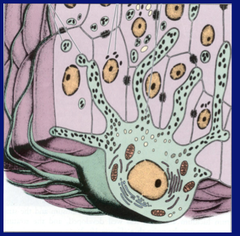
Melanocytes

answer
Present in stratum basale ?Attach to basal lamine w/ hemidesmosomes Main function is to produce melanin, which absorbs UV radiation and protects genetic material of the keratinocytes Extends finger-like processes into the stratum spinosum
question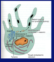
Melanin Production

answer
Produced in melanocytes and is transported into keratinocytes and placed supranuclearly to protect genetic content of cells from UV insult
question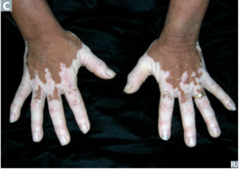
Vitiligo

answer
Hypopigmented/depigmented areas of skin Histopathology: localized loss of melanocytes
question
Albinism
answer
Extremely light complexion Photophobia (redlight reflex in eyes) Photosensitivity Histopathology: no melanin production, tyrosinase missing or mutated
question
Basal Cell Carcinoma
answer
Epithelial tumor Tumor cell nests w/in epidermis and dermis Variable number of melanocytes in tumor Almost never metastasizes so it is different from melanoma
question
Overview of Dermis
answer
Connective tissue ?Dermal papillae (when dermis invaginates into epidermis) ?Epidermal ridges (epidermis invaginates into dermis) Papillary layer Reticular layer Epidermal derivatives invaginate into dermis and multiply ?Hair follicles ?Sweat and sebaceous glands
question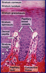
Papillary Layer of Dermis

answer
Loose connective tissue right underneath epithelium Anchoring collagen fibrils in reticular lamina Most easily seen in dermal papilla
question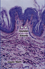
Reticular Layer of Dermis

answer
Darker, more fibrous Dense, irregular connective tissue Elastic fibers Less cellular Makes up majority of dermis
question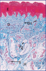
Hypodermis

answer
Loose connective tissue Superficial fascia of gross anatomy Not a layer of the integument
question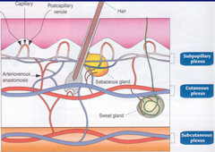
Thermoregulation

answer
Have arterial and venous plexuses throughout skin ?Venous helps to cool off arterial Plexuses exist b/w papillary and reticular layers, between dermis and subcutaneous layers, and middle of the dermis (venous plexus only)
question
Sensory Receptors

answer
Mechanoreceptors Thermoreceptors Nociceptors (pain) Free nerve endings ?Merkels' discs ? responds to mechanical impulses Expanded Ruffini endings ? respond to change in keratin fiber shape Encapsulated Endings ? nerve endings encapsulated in cells, mainly pressure sensors ?Pacinian corpuscle ?Meissner corpuscle ?Krause Corpuscle Peritrichial Nerve Ending ?One nerve for each hair follicle
question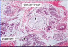
Pacinian Corpuscle

answer
Looks like an onion Deep pressure receptors in hypodermis ?Respond to integumentary movement
question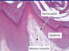
Meissner's Corpuscle

answer
In dermal papilla For fine touch and pressure
question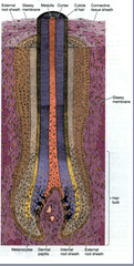
Hair Follicle

answer
Hair bulb ? dermal papillae Hair root Hair shaft ?Medulla, cortex, cuticle, internal root sheath, external root sheath, glassy membrane Extends to outer surface of integument from hypodermis Found in thin skin Have arrector pili muscle which allows hair to stand up
question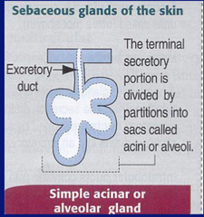
Sebaceous Glands

answer
Acini contain basal layer of undifferentiated epithelial cells Produce sebum Holocrine glands
question
Sweat Glands
answer
Merocrine (produce sweat that goes into center of gland and then out through duct, without losing its shape) or apocrine (very big, secondary sex characteristics so in axilla and groin, lose surface when they excrete) glands Dark and clear cells Myoepithelial cells Stratified cuboidal epithelium
question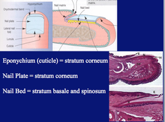
Nail Structure

answer
Nail root - lying in the nail groove Nail plate - rests on the nail bed, stratum corneum Nail bed - regenerative layer, stratum basale and spinosum Eponychium (cuticle) = stratum corneum
question
Evaluating a Skull Series
answer
Look for symmetry Identify the midline Compare paired structures Notice different densities Look for calcifications Observe normal anatomic structures
question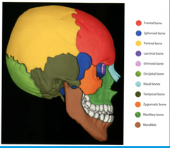
Skull Bones

answer
Calvarium - brain case - skull vault and base Facial bones Mandible Paired parietal bones ?Joined at the midline by sagittal suture ?Form the sides and roof of the cranium Frontal bone ?Forms the front of the skull ?Joined by metopic suture ?Joins the parietal bones at the coronal suture Occipital bone ?Forms the back of the skull ?Joined to the parietal bones at lambdoid suture ?Contains foramen magnum which connects cranial cavity with spinal canal Temporal bone ?At base of skull ?Contains 5 parts ? squama, petrous, mastoid, tympanic parts, and styloid process Sphenoid bone ?At base of skull ?Lesser wings (anterior) ?Sella turcica (pit fossa) ?Greater wings (posterior) Ethmoid bone ?Separates nasal cavity from brain ?Consists of cribriform and perpendicular plates and lateral masses
question
Skull Vault
answer
Composed of flat bones joined at sutures ?Outer table - cortical bone ?Diploic space - cancellous layer with vascular spaces ?Inner table - cortical bone Skull is covered by periosteum which is contiguous with fibrous tissue in sutures
question
Neonatal Skull
answer
Moulding ?Overlapping of cranial bones during birth, resolves in a matter of days Undeveloped Features ?Diploic space ?Vascular markings ?Sinuses not aerated Sutures are straight lines and after 2 years begin to have a serrated border Open fontanelles and close by 15-18 months Skull vault is 8 times the size of facial bones
question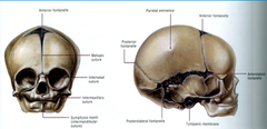
Skull Growth

answer
Fastest during first year of life By age 7 it is nearly adult size Facial bones grow faster than the skull vault Sutures are fused by the second decade Complete bony fusion by the third decade
question
Differentiating Linear Skull Markings
answer
Sutures ?Visible throughout life ?Serpiginous ?White margins Fractures ?More linear ?Not marginated ?More radiolucent (black) than a suture line Vascular markings (grooves) ?Not as radiolucent (black) as fracture lines ?In certain expected locations ?Slightly serpiginous
question
Sutures of Skull
answer
Sagittal - along midline, b/w parietal bones Coronal - b/w frontal and parietal bones Lambdoid - b/w parietal and occipital bones Squamosal - b/w parietal and temporal bones Metopic - b/w 2 frontal bones, prior to fusion
question
Suture Intersections
answer
Bregma - junction of coronal and sagittal sutures Lambda - junction of lambdoid and sagittal sutures Pterion - junction of frontal, sphenoidal, parietal and temporal bones Asterion - squamosal suture meets lambdoid suture
question
Skull Fractures
answer
Classified according to location and type Location: ?Calvaria ?Facial structures ?Base of skull Type: ?Penetrating fractures ?Depressed fractures ?Diastatic fractures (involving sutures)
question
Skull Calcifications
answer
Pineal gland - midline, 50% in adults over 20 y/o Choroid plexus - in bilateral atria of lateral ventricles Dural calcifications Petro and interclinoid ligaments Arachmoid granulations - along the superior midline aspect of the vault Basal ganglia Dentate nucleus Internal carotid artery Lens of eye
question
Function of the Nervous System
answer
Somatic Nervous System ?Motor (efferent) ?Sensory (afferent) Autonomic Nervous System ?Visceral motor (efferent) ?Sympathetic ?Parasympathetic (from cranial or sacral origin) ?Enteric (communicates with CNS through either sympathetic via sympathetic trunks or parasympathetic via vagus) ???Can function entirely by itself, controls peristalsis of stomach ?Visceral sensory (afferent) ?Leads to referred pain Special senses involve specialized organs (i.e. eyes, hair, etc.) ?Include vision, hearing, balance, taste and smell ?Carried to brain by cranial nerves
question
Cell Types in Nervous System
answer
Neurons or nerve cells ?Sensory: somatic, visceral and special senses (vision, hearing, balance, taste and smell) ?Motor: somatic, visceral ?Interneuron ? take up majority of CNS neurons ?Normally connect sensory, motor to form reflex arc Supporting Cells ?Peripheral glial cells ?Schwann cells - produces myelin ???One cell can only produce myelin for one neuron ?Satellite cells ?Central glial cells ?Oligodendrocytes - produces myelin ???Each cell can produce myelin for multiple cells ?Astrocytes ?Microglial cells ?Ependymal cells
question
Neurons

answer
Non-dividing cells (except in hippocampus and olfactory bulbs) Structurally, consist of cell body, dendrites, and axon Has Nissl bodies ? large number in a neuron means that neuron is highly active in protein synthesis ?Stained ribosomes Axon hillock is region in cytoplasm that lacks large organelles, nissl bodies Initial segment of axon is area b/w myelin and axon hillock ?Where AP generates
question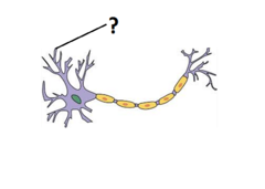
Dendrites

answer
Vary greatly in number and distribution ?Up to 200,000 per neuron Become thinner w/ branching Cytoplasm similar to perinuclear cytoplasm (cytoplasm of soma) except Golgi apparatus
question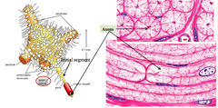
Axons

answer
Transmit information from cell body to other cells Begins at the axon hillock Vary in length and diameter by types ?Usually very long >40in Constant diameter Some are myelinated ?Nodes of Ranvier allow AP to skip over parts of the axon faster
question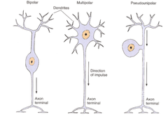
Neuron Morphological Types

answer
Bipolar: special senses Multipolar: motoneurons and interneurons Pseudounipolar: sensory neurons in ganglia ?i.e. dorsal root ganglia, cranial nerve ganglia
question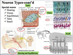
Neuron Types

answer
Somatic Sensory (afferent) ?Location of sensors: Skin, muscles, joints Visceral Sensory (afferent) ?Location of sensors: Internal organs, vessels, glands, mucous membrane Types of sensors ?Nociception - pain ?Thermorepcetion - temperature ?Mechanoreception - movement Special senses ?Hearing ?Taste - bipolar neurons in taste buds ?Vision - bipolar neurons in photoreceptors ?Balance - semicircular canal in inner ear, cochlea ?Smell - bipolar neurons in nasal cavity, olfactory nerve cells Somatic motor (efferent) ?Targets: skeletal muscle fibers ?Forms special synapse with muscle: neuromuscular junction Visceral motor (efferent) ?Targets: smooth muscles, cardiac conducting fibers, glands ?Sympathetic: fight or flight ?Presynaptic: thoracolumbar (IML of T1-L2) ?Postsynaptic: paravertebral and prevertebral ganglia ?Parasympathetic: rest and digest ?Presynaptic: cranial (brainstem) and sacral (S2-4) ?Postsynaptic: parasympathetic ganglia ?Enteric: neurons and their proceeses in walls of GI tract Interneurons ?99.9% of neurons ? most of neurons in CNS ?b/w sensory and motor neurons
question
Synapse
answer
Functional contact b/w neurons or b/w neurons and effector cells (i.e. muscle, gland, etc.)
question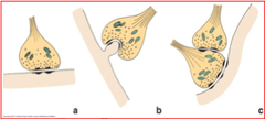
Types of Synapse

answer
Axosomatic = axon on cell body (figure a) Axodendritic = axon on dendrite (figure b) Axoaxonic = axon on axon (figure c) Chemical synapse (neurotransmitters) - have "gain" Electrical synapse (ions): faster conduction, often bi-direction
question
Chemical Synapse
answer
Presynaptic component, synaptic cleft, and postsynaptic membrane Two steps ?Arrival of AP ?Presence of NTs Removal of NT ?Re-uptake into pre-synaptic neuron ?Enzymatic degradation ?diffusion
question
Neurotransmitters
answer
Can be excitatory or inhibitory Classification ?AAs: glutamate, glycine, GABA, etc. ?Monoamines: norepinephrine, epinephrine, dopamine, serotonin, etc. ?Others: ACh, peptides, gases
question
Disorders of Neuromuscular Transmission
answer
Interrupt presynaptic release of ACh (i.e. botulism, Lambert-Eaton syndrome) ?Botulism - attacks proteins associated with synaptic vesicle release of Ach, causing muscle paralysis/weakness ?Lambert-Eaton syndrome - often associated with small cell lung cancer, w/ weakness of some muscles ? auto-antibodies to attack calcium channels, disrupting Ach release Block postsynaptic receptors (e.g. myasthenia gravis) ?Myasthenia gravis - autoimmune disease where patients develop antibody against nicotinic receptor, see muscle weakness Too much ACh depolarize postsynaptic receptors (i.e. cholingeric drugs, organophosphate insecticides and nerve gas) ?Disrupts enzyme (ACh transferase) that breaks down ACh, so it stays in synaptic cleft and continually binds to receptors ? causes muscle convulsions
question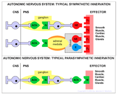
NTs in the ANS

answer
Special case for the sympathetic ? preganglionic neuron synapses directly on adrenal medulla ?Adrenal medulla causes release of NE and epi into blood, causing fight or flight response In the sympathetic, tend to release NE to act on adrenergic receptors ?causes fight or flight In parasympathetic, tend to release ACh to act on musculorinic ACh receptors ?Causes rest and digest response Sweat glands are an exception ?Only receive sympathetic innervation, but follows the parasympathetic pathway (in terms of receptors, use of ACh)
question
Central Nervous System
answer
Includes cerebrum, cerebellum, brain stem, spinal cord Brain stem consists of midbrain, pons, medulla oblongata
question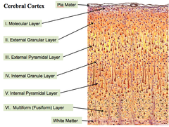
Cerebral Cortex (Transverse Section)

answer
Pia mater is most superficial, white matter is most deep Not just a stack of layers ? function differently Layers 1-3 are mainly for interhemispheric connection (L-R connections) 4th layer are for intrahemispheric connection (w/in one hemisphere) and thalamocortical connections 5th layer contains large pyramidal cells, sending connections to spinal cord, brain stem
question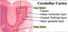
Cerebellar Cortex

answer
3 layers Outer: molecular layer (very light, very few cells) ?Receives axons from granular layer, and dendritic tree from Purkinje layer Central: Purkinje layer ?Very big cells ?Form dendritic trees that send out information Inner: granular layer ?A lot of small cells, creating thick layer ?Axons project to molecular layer
question
Spinal Cord
answer
Dorsal horn - sensory neurons Ventral horn = motor neurons Interneurons lie in between, connecting sensory and motor
question
Central neuroglia

answer
Astrocytes ?2 morphologies: fibrous (white) and protoplasmic (gray) ?express GFAP (glial fibrillary acidic protein) ?maintain blood-brain barrier ?move metabolites to and from neurons ?modulate neuronal survival and activity Oligodendrocytes ?Produce CNS myelin ?One to several neurons Microglia ?Phagocytic cells ?When activated, retract processes and appear like macrophages ?Involved in inflammation and repair Ependymal Cells ?Epithelium-like cells lining ventricles of brain and central canal of spinal cord ?Cilia and microvilli on apical (facing center of canal) surfaces ?Choroid plexus (modified ependymal cells + associated capillaries) ?Produce CSF and have microvilli for filtration
question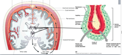
CSF Production and Flow

answer
CSF is produced in choroid plexus lining the 4 ventricles Flows from 4th ventricle into subarachnoid space (through median and lateral apertures) Small amount flows into central canal of spinal cord CSF can be reabsorbed CSF has poor protein level but high glucose level (compared to blood)
question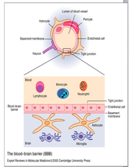
Blood-Brain Barrier

answer
Anatomy ?Tight junctions of endothelial cells, lack of fenestration ?Basement membrane ?Astrocyte end feet ?pericytes Function ?Filter and restrict passage of certain molecules (>500 daltons) ?Maintain water and ion levels in the CNS ?Blood-borne immune cells cannot penetrate (e.g. lymphocytes, monocytes, and neutrophils)
question
Diseases involving the BBB
answer
?Meningitis ?MS ?Alzheimer's ?HIV encephalitis ?Rabies
question
Peripheral Nervous System
answer
Nerves ?Cranial and spinal nerves ?Somatic branches ?Autonomic branches ?Parasympathetic ?sympathetic Ganglia ?Sensory ganglia ?Autonomic ganglia ?Parasympathetic ?Sympathetic
question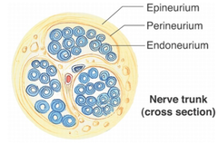
Nerve coatings

answer
Epineurium ?Membrane that surrounds entire nerve Perineurium Endoneurium ?Very thin, wraps individual neuron
question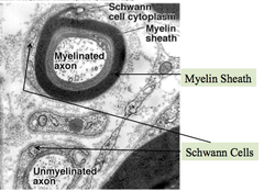
Peripheral Glial Cells

answer
Schwann cell ?Supported myelinated and unmyelinated axons ?Produce myelins to speed up conduction of nerve impulses ?Can only myelinate one axon ?Can provide more than one myelin per axon ?Clean up debris and guide regeneration Satellite cells ?Electric insulation ?Metabolic exchange
question
Eye Development

answer
Primary induction - notochordal process and notochord induce overlying ectoderm to form forebrain Secondary induction sequence ?Mesoderm adjacent to forebrain induces it to form an optic vesicle ?Optic vesicles induce overlying ectoderm to form lens placodes which will form the lenses ?Lens epidermis induces overlying ectoderm to form transparent cornea
question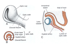
Optic Cup

answer
Origin: begins to form on day 22 Development ?Evagination of the wall of forebrain projects into adjacent mesoderm as optic vesicle ?Vesicle then invaginates distally to form double-layered optic cup while proximal end constricts to form optic stalk ?Optic (choroid) fissure forms in inferior surface of optic cup and optic stalk and mesoderm which invades this fissure gives rise to hyaloid blood vessels which will supply inner eye (lens vesicle, optic cup) ?When optic fissure closes, blood vessels become trapped w/in optic nerve Forms retina, ciliary body and iris
question
Retina Development

answer
Develops from posterior 2/3 of the optic cup Inner thick layer of cup becomes neural retina Optic nerve is formed by axons sent into optic stalk by ganglion cells in the neural retina ?Note that the optic nerve is not a true cranial nerve but part of the brain since both the neural retina and optic stalk are derivatives of the forebrain ?Consequences of this are enormous since meningeal coverings and spaces b/w each of the brain are the coverings/spaces around optic nerves ?Ophthalmologic examination therefore provides not only information about the eye but also a window on conditions w/in cranial cavity and membranes/spaces around brain Outer thin layer of cup becomes pigmented layer of retina Two retinal layers fuse during fetal period but pigmented layer adheres more strongly to choroid than it does to neural retinal layer so that the neural retina is easily detached (congenital detachment of the retina) Completely formed w/in 3-4 months postnatally (only fovea not formed at birth)
question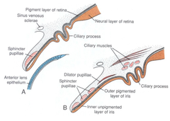
Ciliary Body ; Iris Development

answer
Develop from anterior 1/3 of optic cup Ciliary body ?Both layers of optic cup contribute to its formation ?Pigmented part of body is rom outer layer and non-pigmented part is from the inner layer (contains no neural elements) ?Mesoderm at the edges of optic cup gives rise to ciliary muscle and connective tissue of ciliary body Iris ?Represents the forward extension of the ciliary body over the edges of the lens ?Non-pigmented at birth (light blue or gray) and becomes blue (pigment in outer layer of the iris only) or brown pigment in outer layer of iris and surrounding connective tissue at 6-10 months ?Dilator and sphincter muscles are derived from neural crest cells in the choroid and NOT from mesoderm ?Blood supply to iris is from outside the eye via anterior ciliary arts from the ophthalmic artery
question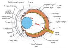
Lens Development

answer
Origin: begin on day 28 Development ?Begins as a lens placode in surface ectoderm overlying optic vesicle ?Placode invaginates to form lens vesicle ?This vesicle is invested by mesoderm which forms a vascular layer around the lens (tunica vascualris lentis) supplied by the hyaloid artery ?Wall of vesicle forms a cuboidal lens epithelium anteriorly and posteriorly the cells elongate into lens fibers ?These fibers grow into and occlude the cavity of the lens vesicle ?New fibers are added continuously at the lateral edge of the equatorial zone (until about 20 years of age) and these fibers grow towards both the anterior and posterior poles of the lens ?As the fibers mature, the nuclei die and the fibers become transparent ?Epithelial cells making up the wall of the lens vesicle also secrete an elastic material which covers the lens as the lens capsule
question
Vitreous Body Development
answer
Origin: secreted during 2nd month (late embryonic stages) Development ?Inner cell layer of optic cup (neural retinal layer) secretes a translucent ground substance containing a delicate meshwork of fibers ?Invading mesodermal cells also contribute to this fiber formation ?A gelatinous transparent material is then laid down between these fiber and together this material and the fibers make up the vitreous body
question
Aqueous Chamber ; Cornea Development
answer
Origin: 6th-7th week Development ?Aqueous chamber forms from 2 mesodermal cavities ?Mesoderm lying b/w lens and ectoderm cavitates to form the anterior chambers ?Mesoderm lying anterior to this chamber form the substantia propria of the cornea and the mesothelium of the anterior chamber ?The mesoderm lying posterior to chamber is applied to surface of the lens and iris and forms the pupillary membrane ?Mesoderm lying b/w inner surface of iris and lens then cavitates to form the posterior chamber ?Anterior and posterior chambers remain separated by pupillary membrane ?These two chambers form the aqueous chamber when the pupillary membrane degenerates late in fetal life ?Aqueous humor is secreted by cells in the ciliary process into aqueous chamber ?Fluid is reabsorbed by sinus venosus sclerae (canal of Schlemm) ?Degeneration of pupillary membrane and tunica vascularis lentis overlying the anterior part of the lens results from degeneration of the terminal branches of the hyaloid vessels which supplied these structures ?Traces of these degenerated hyaloid vessels can be seen as a hyaloid (Cloquet's) canal through the vitreous body ?Proximal parts of hyaloid vessels remain to become central vessels of retina ?Cornea is composed of both mesoderm (substantia propria) and overlying ectoderm
question
Sclera & Choroid Development
answer
Origin: 6th week Development ?Mesoderm and neural crest surrounding the optic cup differentiates into an inner vascular choroidal layer and an outer fibrous scleral layer ?Sclera continues over eye anteriorly as substantia propria of the cornea NOTE: choroid and sclera are continuous w/ coverings of brain ?Pia-arachnoid of brain is continuous with the choroid layer (vascular) and the sclera with the dura mater ?Subarachnoid space continues down over the optic stalk (nerve) from the brain but ends at the eyeball where the sclera and choroid are tightly fused together
question
Eye Muscle Development
answer
Probably arise from pre-otic somites Although pre-otic somites are not seen in man they are present in lower animal forms such as sharks
question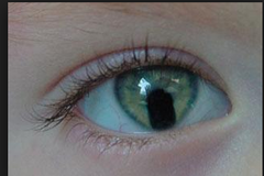
Coloboma Iridis

answer
Results from failure of optic fissure to close during 7th week Defect is usually confined to the iris giving the pupil a keyhole appearance but it can include the retina, choroid and optic nerve
question
Persistent Pupillary Membrane
answer
Results from the failure of the hyaloid vessels to degenerate completely so that the pupillary membrane over the lens persists Occurs during late fetal life and usually has little effect on vision
question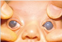
Congenital Glaucoma

answer
Aka buphthalmos Results from failure of proper drainage system (sinus venosus sclerae) to form for the aqueous humor Intraocular pressure damages the retina and swells the eyeball This defect occurs in 0.5% of births and can be caused by rubella infections or a recessive genetic disorder
question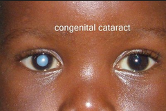
Congenital Cataract

answer
Results from either the failure of the lens to clear or re-clouding of the lens secondary to infection Occurs during 4th-8th week and may be genetic or caused by rubella infection
question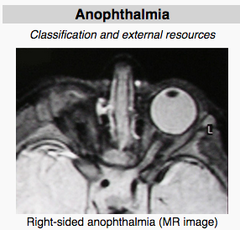
Microphthalmia and Anophthalmia

answer
Results either from failure of optic vesicle to form (anopthalmia) or from abnormal optic cup formation from the optic vesicle (microphthalmia) In anophthalmia, neither lens, retina nor cornea are present In microphthalmia, the eye is small to rudimentary ?Lens and cornea may be missing but retina is present although it may be rudimentary) Occurs during 4th week
question
Cyclopia
answer
When the prechordal plate mesoderm (midline mesoderm just caudal to oropharyngeal membrane) fails to induce normal forebrain formation in the cranial neural plate, the undervelopmed forebrain causes the optic vesicles to grow in a cranial direction so that they are very close together or fuse to form one optic vesicle 1/40,000 births defects ranges from 1 median eye (true cyclopia) to 2 eyes in one orbit, 2 eyes in separate orbits very close together
question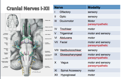
Cranial Nerves I-XII

answer
There are 4 spinal nerves that have parasympathetic function ? basically lead to secretions
question
Modalities of Cranial Nerves
answer
Sensory modalities ?General sensory - touch, pain, temp, pressure/vibration, proprioception ?Visceral sensory - sensory input from viscera ?Special sensory - smell, vision, taste, hearing and balance Motor modalities ?Somatic motor - muscles that develop from somites ?Branchial motor - muscles that develop from branchial arches ?Visceral motor - innervates viscera, including glands and all smooth muscle
question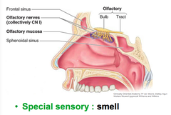
Olfactory Nerve (CN I)

answer
Pokes through cribriform plate ?Nerve projects directly into cerebral cortex ?Primary cell bodies poke through the cribriform plate Special sensory: smell Both sides of CN I connect to each other so that we smell with each nostril ?Connect at the anterior commissure
question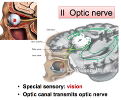
Optic Nerve (CN II)

answer
Special sensory: vision Attaches to posterior region of eye Optic canal transmits optic nerve Optic chiasm crosses over, sending fibers from one side to the other ?Splits axonal fibers b/w brain hemispheres
question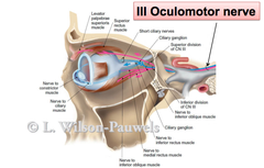
Oculomotor Nerve (CN III)

answer
Somatic motor ?Eye movement (4 of the 6 extrinsic muscles for eye movement) ?Medial rectus ?Inferior rectus ?Superior rectus ?Inferior oblique ?Maintains an open eyelid (via levator palpebrae superioris) Visceral motor (parasympathetic) ?Control size of pupil and shape of lens for accommodation of vision ?Innervates constrictor pupillae muscle and ciliary body for accommodation ?Ciliary ganglion is the major cel body associated with parasympathetic component here ?Sends branches via short ciliary nerves ?Innervates ciliary body for accommodation ?Hitches a ride w/ nasociliary branch of trigeminal nerve
question
Accommodation Reflex
answer
Adaption of the visual apparatus of eye for near vision Eyes converge ? medial rectus innervated by CN III Then the sphincter pupillary muscle in iris and the ciliary muscles contract ?Sphincter pupillary muscle in iris constricts ? pupils constrict ?Ciliary muscle contract - decreased tension on suspensory ligament, leading to a thicker lens As we age, our lens starts to thicken ?makes it more difficult for suspensory ligaments to tug and release lens, so we end up with poor near vision
question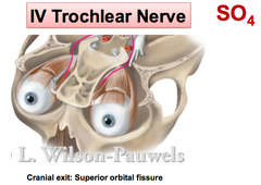
Trochlear Nerve (CN IV)

answer
Cranial exit: superior orbital fissure Somatic motor: innervates superior oblique muscle of the eye ?"downward gaze"
question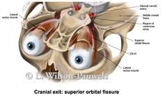
Abducens Nerve (CN VI)

answer
Somatic motor: innervates lacteral rectus, only muscle that can abduct your eye ?"lateral gaze"
question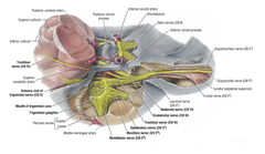
Trigeminal Nerve

answer
Gives off three branches: ophthalmic, maxillary, mandibular ?Each of these branches has many further branches Major sensory cranial nerve of the face and head Control muscles involved in chewing General sensory: V1, V2, V3 ?Outside cranium: face, nasal cavity, oral cavity ?Inside cranium: meninges Brachial motor: V3 ?Muscles of mastication (masseter, temporal, medial and lateral pterygoid) Cranial exits: ?V1 - superior orbital fissure ?V2 - foramen rotundum ?V3 - foramen ovale
question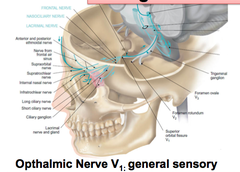
Ophthalmic Nerve (V1)

answer
General sensory of the orbit Supplies cornea, superior conjunctiva, mucosa of anterosuperior nasal cavity, frontal/ethmoidal/sphenoidal siuses, anterior and supratentorial dura mater, skin of dorsum of external nose, superior eyelind, forehead and anterior scalp Also innervates part of the meninges
question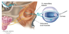
Ophthalmic Nerve Blink Reflex

answer
Touch cornea w/ cotton swab If you do it with one eye, the other eye should blink too
question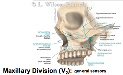
Maxillary Division (V2)

answer
General sensory Supplies dura mater of anterior part of middle cranial fossa, conjunctiva of inferior eyelid, mucosa of postero-inferior nasal cavity, maxillary sinus, palate and anterior part of superior oral vestibule, maxillary teeth, and skin of lateral external nose, inferior eyelid, anterior cheek and upper lip Need to know superior alveolar nerves (major branch of V2) ?Innervate upper row of teeth
question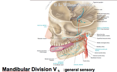
Mandibular Division (V3)

answer
Innervates lower teeth and jaw, anterior 2/3 of tongue ?Has inferior alveolar nerves as a major branch General sensory innervation of tongue ? NOT TASTE Branchial motor division ?Supplies motor innervation to 4 muscles of mastication, mylohyoid, anterior belly of digastric, tensor veli palatine, and tensor tympani
question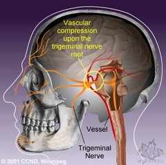
Trigeminal Neuralgia

answer
Tic douloureux (painful spasm) Generally affects maxillary-mandibular branch Pain is lancinating (shock like pains) and lasts a few seconds to a few minutes ?Can last days, weeks or months on end and then not return for months of even years Occurs spontaneously or elicited by eating, talking, shaving, brushing teeth (touching a trigger zone) Occurs most often in people over age 50, but it can occur at any age Disorder is more common in women than in men Is incredibly difficult to diagnose and to treat
question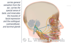
Facial Nerve (CN VII)

answer
Carries general sensation from ear, carries special sense of taste, innervates muscles of facial expression and the sublingual, submandibular and lacrimal glands Major components ?Branchial "somatic" motor: muscles of facial expression ?Visceral motor (parasympathetic) - lacrimal, submandibular/sublingual glands, mucous membranes of nasopharynx and palate ?General sensory - skin of external acoustic meatus and external tympanic membrane ?Special sensory - taste anterior 2/3 of tongue ?Chorda tympani ? for taste, associated with geniculate ganglion Cranial exits: internal acoustic meatus
question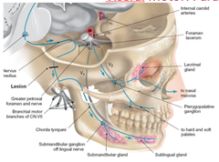
Parasympathetics of Facial Nerve

answer
Stimulations: ?Lacrimal gland ?Mucus membrane of nose ?Submandibular gland ?Sublingual gland Pterygopalatine ganglion - great petrosal nerve ?Hang onto V2 branch Submandibular ganglion - chorda tympani ?Hang onto V3 branch
question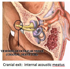
Vestibulocochlear Nerve (CN VIII)

answer
Special sensory: Involved in both hearing and balance ?Balance portion is extremely complicated Cranial exit: internal acoustic meatus
question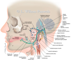
Glossopharyngeal Nerve (CN IX)

answer
Major components ?Branchial motor: stylopharyngeus muscle ?Special sensory: taste posterior 1/3 tongue ?General sensory: external ear, posterior 1/3 of tongue ?Visceral sensory: parotid gland, pharynx, middle ear, carotid body, carotid sinus ?"gag reflex" ?visceral motor: parotid gland (parasympathetic) ?lesser petrosal nerve synapses on otic ganglion - parotid gland ("hitch a ride" on V3) Cranial exit: jugular foramen
question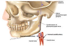
Visceral Sensory Function of the Glossopharyngeal Nerve

answer
Visceral sensory: subconscious sensation Carotid body (chemoreceptors) ? monitors oxygen tension in blood, carbon dioxide, and acidity/alkalinity Carotid sinus (baroreceptors) - monitors arterial blood pressure
question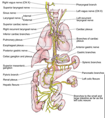
Vagus Nerve (CN X)

answer
"from the brain stem to the splenic flexure of the colon" Motor: palate, pharynx, larynx, tracheobronchial tree, heart, GI tract to the left colic fixture ?Cardiac: slows HR ?Lungs: stimulates increased bronchiolar secretions and bronchoconstriction ?GI tract - stimulates increased secretions and motility Sensory: pharynx, larynx, reflex sensory from tracheobronchial tree, lungs, heart, GI tract to left colic fixture Visceral Sensory ?Aortic arch (baroreceptors) ?Stretch receptors that monitor BP ?Aortic bodies (chemoreceptors) ?Measures oxygen tension in blood Visceral motor (parasympathetic) ?Smooth muscle of trachea, bronchi, digestive tract ?Moderates cardiac pacemaker and vasoconstrictor of coronary arteries Recurrent laryngeal nerves are branches of vagus that supply larynx (except cricothyroid muscles) ?Emerges from vagus at level of arch of aorta Cranial exit: jugular foramen
question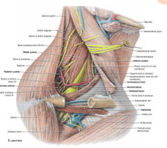
Spinal Accessory Nerve (CN XI)

answer
Somatic motor: sternomastoid muscle, trapezius muscle Cranial exit: jugular foramen
question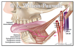
Hypoglossal Nerve (CN XII)

answer
Innervates extrinsic and intrinsic muscles of the tongue ?Allows you to stick you tongue out, lick things Cranial exit: hypoglossal canal Genioglossus (testing for lesion) ?Ask patient to stick their tongue out straight
question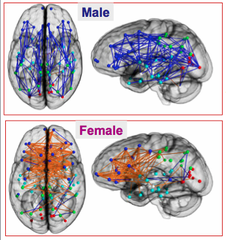
Are male and female brains different?

answer
Male brains are structured to facilitate connectivity b/w perception and coordinated action Female brains are designed to facilitate communication between analytical and intuitive processing modes
question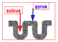
Basic Construct of Cerebrum

answer
Gyrus - folds or ridge in cerebral cortex, surface b/w an adjacent sulcus Sulcus - groove b/w gyrus Fissures - deeper sulcus
question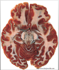
Gray and White Matter in the Cerebrum

answer
Gray matter = neural cell bodies White = myelinated axonal fibers The surface of the cerebrum is comprised of numerous folds or convolutions of gray matters (neurons) and underlying white matter (axons)
question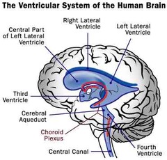
Ventricular System

answer
Filled w/ CSF Consists of lateral ventricles, 3rd ventricle and 4th ventricle Choroid plexus secretes CSF Key to pathology associated w/ the brain
question
Subcortical Structures
answer
Diencephalon ?Thalamus (relay sensory and motor) ?Hypothalamus (link to endocrine system) Limbic (emotions and memory) ?Hippocampus ?Amygdala ?Olfactory bulb Basal ganglia (movement control) ?Caudate nucleus ?Putamen ?Globus pallidus ?Accumbens nucleus
question
Brainstem
answer
Consists of the midbrain, pons, and medulla Regulatory structures from breathing and HR are located here Functions ?Conduit for ascending or descending fiber tracts ?Cranial nerve nuclei ?integration
question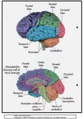
Organization of Cerebral Cortex

answer
Central sulcus divides the frontal lobe ?Frontal lobe is motor cortex Posterior to frontal is the parietal lobe ?Controls all things somatosensory Occipital lobe is most posterior ?Integration Temporal lobe is most lateral ?Important for auditory information Limbic lobe is where the hippocampus and amygdala are housed for memory and emotional responses Insular lobe is important for taste, pain (and audition, secondarily)
question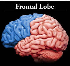
Frontal Lobe

answer
Planning and organization Problem solving and decision making Expressive language Controlling behavior, emotions and impulses ?Cross-talks w/ limbic limb Motor execution
question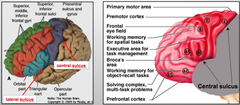
Frontal Lobe Subdivisions - Motor Cortex

answer
BA 4 - Primary motor cortex ?Precentral gyrus BA 6 - Premotor cortex ?Superior and middle frontal gyrus, precentral gyrus BA 8 - Frontal eye field ?Superior frontal (middle) gyrus BA 44/45 - Broca's area ?Speech production ?Inferior frontal gyrus
question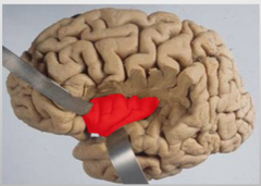
Insular Cortex

answer
Integrates pain Also has role in taste, emotion and cognition BA 16 - located deep within lateral sulcus, deep to frontal cortex
question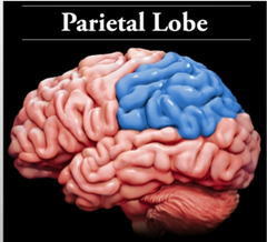
Parietal Lobe

answer
integrate sensory information from various parts of the body Spatial reasoning contains the primary sensory cortex, which controls sensation ?touch, hot or cold, pain, pressure
question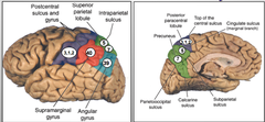
Parietal Lobe Subdivisions - Somatosensory Cortex

answer
BA 3, 1, 2 - Primary somatosensory cortex ?Postcentral gyrus BA 5, 7 - somatosensory association cortex ?Superior parietal lobule ?Involved in spatial perception BA 39 - recognition of visual symbols ?Angular syrus ?i.e. seeing stop sign and knowing what it is w/o reading it BA 40 - integrates auditory commands ?Supramarginal gyrus ?Not a primary auditory cortex
question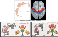
Homonculus: Somatotopic Mapping

answer
We know specifically when we talk about primary motor and sensory cortex where the innervation is coming from and going to from primary cortices ?Medially ? genitalia and extremities ?Laterally ? face, eyes, tongue, etc.
question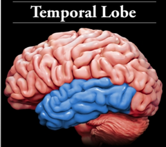
Temporal Lobe

answer
Recognizing and processing sound Understanding and producing speech Various aspects of memory
question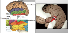
Temporal Lobe Subdivision - Auditory Cortex

answer
BA 22 - Wernicke's area ?Where comprehension of speech actually occurs ?Superior temporal gyrus (posterior portion) BA 41 - Primary auditory cortex ?transverse temporal gyrus (of Heschl) BA 42 - auditory association cortex ?Transverse temporal gyrus
question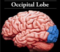
Occipital Lobe

answer
Receive and process visual information Contain areas that help in perceiving shapes and colors
question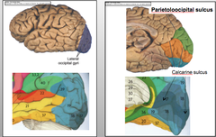
Occipital Lobe Subdivisions - Visual Cortex

answer
BA 17 - Primary visual cortex ?Banks of calcarine sulcus ?A stroke here will compromise any kind of visual processing BA 18, 19 - visual association area ?Surrounding BA 17
question
Cortical Connections
answer
Projection fibers connect cerebral cortex w/ subcortical nuclei, brainstem and spinal cord Commissural fibers connect cortex b/w right and left cerebral hemispheres Association fibers connect regions of cerebral cortex w/in the same hemisphere
question
Projection fibers
answer
Projection fibers connect cerebral cortex w/ subcortical nuclei, brainstem and spinal cord Stays highly organized from cerebrum through projections in spinal cord ?Allows mapping different regions that it targets throughout body
question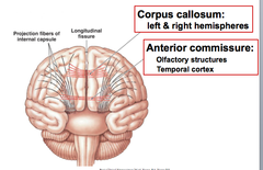
Commissural Fibers

answer
Commissural fibers connect cortex b/w right and left cerebral hemispheres 2 types ?Corpus callosum is a major fiber tract that connects the left and right hemispheres ?The anterior commissure connects the olfactory structures and the temporal cortex ?Much smaller than the corpus callosum
question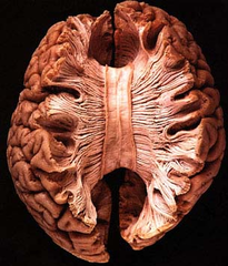
Corpus Callosum

answer
Can take longitudinal fissure and pull it apart to see corpus callosum Consists of genu, body and splenium ?Genu connects frontal lobe structures
question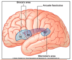
Association fibers

answer
Association fibers connect regions of cerebral cortex w/in the same hemisphere Arcuate fasciculus connects BA 22 (Wernicke's area) to BA 44, 45 (Broca's area) ?Left hemisphere dominance
question
Aphasia
answer
A disturbance in dominant hemisphere that produces a defect in the expression or comprehension of any forms of language Caused by focal brain lesion (stroke, head injury) Left hemisphere in 96% of cases
question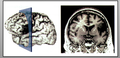
Broca's Aphasia

answer
"expressive" or nonfluent comprehension of language is normal, but unable to convey thought into meaningful language ?inability to organize words into sentences nonfluent speech articulation is impaired BAs 44, 45
question
Wernicke's Aphasia
answer
Fluent Comprehension of language is impaired Fluent speech that is unintelligible Progression of pathology can affect writing, other forms of communication as well BA 22
question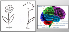
Contralateral Neglect Syndrome

answer
Right parietal cortex damage ? neglect left half of the world ?includes visuospatial tasks, as well as personal neglect
question
Flexibility vs. Stability in the Neck
answer
The neck must be flexible region to allow head to move in many directions, detect threats The downside of flexibility is that to allow flexibility it means that the neck is not a very protected area ?There are many arteries and nerves in this area that are not very protected Stability is sacrificed for flexibility
question
Boundaries of the Neck
answer
Planes are somewhat oblique Posterior - cervical vertebrae Anterior - series of components that make structural component of neck Superior - Hyoid bone, thyroid cartilage, cricoid cartilage and trachea ?Anterior - hyoid bone Inferior - manubrium and clavicles
question
Hyoid Bone
answer
Very protected ?Would probably be protected U-shaped structure w/ body and greater and lesser horn ?Lesser horn is attached to ligament that is attached to styloid process - stylohyoid process ?the one structural attachment to skeleton ?Can ossify as you age ?Body has attachment of a number of muscles that act on thyroid cartilage and hyoid bone Not free floating but also not attached to another bone Hyoid fracture can occur ?Coroners look for in case of strangulation
question
Thyroid Cartilage
answer
Connected to hyoid bone by thryohyoid membrane ?Ligaments here are just thickening of membrane ?Keeps it moving synchronously Sort of two halves that come together to form a laryngeal prominence (Adam's apple) ?More prominent in men ?Moves up and down when speaking Has superior and inferior horn ?Superior horn heads medially and superiorly, w/ attachment to hyothyroid ligament ?Inferior horn connects to cricoid cartilage (located ~C6) ?Cricoid cartilage is a small cartilage that looks like a ring (with large oval part posteriorly) ?b/w thyroid cartilage and cricoid cartilage is the cricothyroid membrane which has thickenings to form cricothyroid ligament ?this membrane also helps to keep these cartilages moving synchronously sitting directly behind this cartilage is the larynx damaging this cartilage has potential to damage vocal cords, impede the airway
question
Trachea
answer
Cartilaginous series of C-shaped rings w/ a deficit posteriorly ?Posteriorly, you have esophagus ?When you swallow and esophagus expands, you don't want it to hit anything hard Posteriorly, trachealis muscles connect the rings, allowing for soft tissue so esophagus can expand This is at risk b/c it is below mandible
question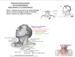
Which zone of the neck is considered the most vulnerable?

answer
Neck is divided into 3 zones Zone 1: clavicle & manibrium up to cricoid cartilage (approximately C6) Zone 2: cricoid cartilage to angle of mandible •Zone 2 is the most accessible Zone 3: superior to angle of mandible Risk for morbidity and mortality is higher in zones 1 and 3 b/c they are much more difficult to access •If you penetrate these zones w/ bullet/knife, it is much more difficult to stop the bleeding
question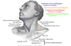
Cervical Regions of the Neck

answer
Posterior cervical region is essentially the trapezius muscle Lateral Cervical Region (posterior triangle) is b/w sternocleidomastoid, trapezius and clavicle Sternocleidomastoid Region is basically the sternocleidomastoid muscle Anterior Cervical Region (anterior triangle) is b/w sternocleidomastoid, mandible, and midline of neck
question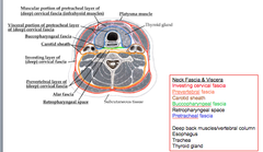
Fascial Planes of the Neck

answer
Deep to superficial fascia, there is an investing cervical fascia ?It is the deep fascia around entire neck ?Runs in and surrounds sternocleidomastoid and trapezius ?Anteriorly, stops at the manubrium Neck is then subdivided again into an anterior and posterior region, w/ some smaller regions as well The posterior region contains the deep muscles of the back around vertebral column and is surrounded by prevertebral layer of fascia The anterior region has a number of viscera including trachea, thyroid gland and esophagus, all of which are surrounded by the pretracheal fascia Laterally, there is another subdivision ? carotid sheath ?Contains carotid artery, internal jugular vein and the vagus nerve
question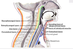
Retropharyngeal space is at risk for infection

answer
Esophagus is continuous with pharynx, which has its own fascia called the buccopharyngeal fascia (continuous with pretracheal fascia) Important b/c space b/w pretracheal fascia and anterior part of the prevertebral fascia is called the retropharyngeal space ?Runs from cranium all the way down to the fibrous pericardium ?Can transmit infection from head to thorax, or vice versa ?Can have pus build up in that space, causing compression of structures
question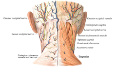
Posterior Cervical Region

answer
Contains the trapezius muscle Place that you palpate in OMM b/c there is a lot of pain in the back and muscles a little deeper
question
Sternocleidomastoid Region
answer
Sternocleidomastoid has 2 heads ?Clavicular head ?Sternal head (attached to manubrium) Innervated by spinal accessory nerve Unilaterally, will flex head to ipsilateral side, rotate to contralateral side Using them together ? extend head at atlanto-occipital joints Torticollis ?Sometimes children born with it, not sure why it occurs ?Suggested that there is not enough blood supply to muscle ?Essentially sternocleidomastoid begins to shorten on one side, pulling and creating action of unilateral contraction ?Have head flexing to one side and rotating to the other, leaving head in this position permanently ?If this is not addressed in development, you can affect formation of skull ?Treatment requires muscle to be severed to allow head to go back to neutral position
question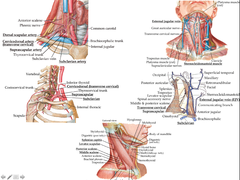
Lateral Cervical Region

answer
Aka posterior triangle Posterior to sternocleidomastoid, superior to clavicle, anterior to trapezius Contains subclavian artery runs posterior to the anterior scalene ?Branches that come off the subclavian come off posterior to the anterior scalene ?Transverse cervical (cervicodorsal) artery and the suprascapular artery Veins of this region ?External jugular vein is very superficial and runs over sternocleidomastoid, then runs deep and empties into subclavian vein ?Formed by posterior auricular vein and the anterior branch of the retromandibular vein ?Subclavian vein ?Central lines usually placed here Muscles in this compartment can help you laterally flex head, anterior flex head (bilaterally) and rotate the head (unilaterally) ?Splenius capitus ?Levator scapulae ?Posterior scalene ?Middle scalene Levator scapula syndrome is caused by excessive rotation or over use of the muscle and they can have crepitus and irritate the muscle, causing radiating pain in neck For a majority of these anterior muscles they are innervated by ventral rami ?Splenius capitus is innervated by dorsal rami ?Levator scapulae is innervated by dorsal scapular nerve
question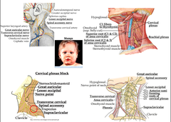
Nerves are the major component of the lateral cervical region

answer
Posterior to the sternocleidomastoid about 1/3 up from the clavicle is a point called the nerve point where many of these nerves exit Great auricular nerve travels superficial to the sternocleidomastoid and up to ear ?Innervates fascia of parotid gland ?This fascia is very dense and doesn't stretch ?In mumps, the parotid gland swells and activates the great auricular nerve, causing pain Lesser occipital nerve provides cutaneous innervation to the back of the head Transverse cervical nerve comes across sternocleidomastoid and supplies cutaneous innervation to anterior neck Supraclavicular nerves travel in an inferior direction and are pretty substantial in size ?Go over should and provide cutaneous innervation to the shoulder All of these nerves are from the cervical plexus (C1-C4) except for the spinal accessory nerve (CN XI) Two more nerves in this region are deep to the muscles ?Ansa cervicalis forms a loop via two roots: superior (C1 and C1) and inferior (C2 and C3) ?This loop forms anterior to carotid sheath ?Phrenic nerve is formed from C3, C4, C5 (need to know these roots) ?Provides sole innervation to diaphragm When you perform surgery in this region, anesthetize the nerve point, which anesthetizes the cervical plexus, which affects part of the phrenic nerve ?If both sides are anesthetized this will paralyze both phrenic nerves and will cause you to not be able to breath w/o a ventilator
question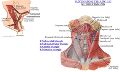
Anterior cervical region

answer
Defined as anterior to sternocleidomastoid, inferior to mandible and along midline Further subdivided into 4 triangles ?Submental ?Submandibular ?Carotid ?muscular
question
Submental triangle
answer
Defined by anterior belly of the digrastic muscle and inferiorly by hyoid bone Not a whole lot goes on here
question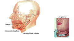
Submandibular Triangle

answer
Defined by anterior and posterior bellies of digastric along w/ mandible Contains submandibular gland Facial layers in oral cavity are somewhat continuous w/ submandibular triangle Infection in mouth can be transmitted to this triangle, space in this region will increase and can push structures into oral cavity Ludwig's angina = cellulitis or infection of fascia in submandibular triangle, causing swelling which pushes up into oral cavity and impacts tongue, pushing it out of mouth ?impacts respiration and swallowing
question
Carotid Triangle
answer
defined by sternocleidomastoid, posterior belly of the diagrastic and omohyoid muscle External carotid artery ?Pulse checked here ?If pulse here is not good an angiogram can be used to check for stenosis Internal jugular vein (IJV) ?Behind sternocleidomastoid ?Continuation of sigmoid sinus, exits jugular foramen and becomes IJV ?As it enters subclavian there is an inferior bulb and valve of IJV (but it is non-functional), but is essentially a valveless vein ?If there is occlusion to this vein, blood would back up into dural venous sinuses and increase intracranial pressure and cause backup of CSF, increasing intracranial pressure Branches of external carotid artery to neck ?Superior thyroid and superior laryngeal ?Lingual ?Facial ?Occipital ?Posterior auricular ?Ascending pharyngeal ?Sometimes the lingual and facial artery come off as a common trunk, but they can also come off separately Internal carotid is NOT part of this triangle ?There is a dilated area of the internal carotid just after it splits from common carotid called the carotid sinus ?This area senses BP via baroreceptors ?On top of this area are chemoreceptors called the carotid body that sense low O2 and high CO2, and regulate accordingly Lymph ?Majority of body lymph drains into thoracic duct, which empties into left subclavian left ?right head and right half of upper head drains into right lymphatic duct ?Empties into right subclavian vein ?Flow from head and neck travels from superior to inferior ?Nodes: ?Submandibular ?Pre-lyngeal ?Infrahyoid ?Pre-tracheal ?supraclavicular
question
Muscular Triangle
answer
Borders are defined by sternocleidomastoid, inferior aspect of omohyoid muscle, and midline of neck Have very large muscular content ?Infrahyoid muscles are contained here Innervation to this triangle is from the ansa cervicalis except the thyrohyoid, which gets its own personal branch from fibers of C1 Major functions of these muscles are to raise and lower hyoid bone and thyroid cartilage ?Muscles are important to strength after an even like a stroke b/c they help in swallowing Superficial muscles in this triangle ?Sternohyoid ?Superior belly of omohyoid connected to hyoid bone ?Superior and inferior omohyoid muscles are connected via an intratendinous section ?Intratendinous section is attached to the clavicle ?Inferior belly of omohyoid is connected to the scapula Deep muscles ?These two muscles look like a deep portion of the stylohyoid ?Thyrohyoid ?This is the superior piece ?From hyoid to thyroid cartilage ?Innervated by fibers from C1 ?Sternothyroid ?This is the inferior piece ?From thyroid cartilage to sternum
question
Suprahyoid Muscles
answer
The suprahyoid muscles are not in muscular triangle, but they attach to the hyoid Elevate the hyoid bone Anterior belly digastric ?From mandible to hyoid ?Innervated by myohyoid nerve Posterior belly digastric ?From hyoid to mastoid process ?Both muscles are connected via an intratendinous sling, which is connected to the hyoid bone itself. ?Innervated by facial nerve Stylohyoid ?From styloid process to hyoid ?The facial nerve passes between the stylohyoid and posterior belly of the digastric. ?Innervated by the facial nerve. Mylohyoid ?Innervated by mylohyoid nerve ?Creates the floor of the mouth. ?Attached to the body of the hyoid bone and to the mylohyoid line along the mandible. Geniohyoid ?Innervated by fibers of C1 ?Superior to the mylohyoid muscle
question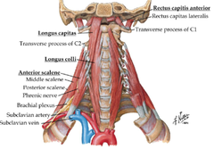
Deep muscles of the neck

answer
Analogous to the transversospinalis muscles of the deep back Found on the deep anterior portion of the neck, running along the vertebral bodies Muscles: ?Rectus capitis anterior ?Long capitas ?Longus colli ?Anterior scalene Innervated by ventral rami Major function of these muscles are lateral flexion, rotation and anterior flexion In whiplash injury, these muscles are potentially damaged
question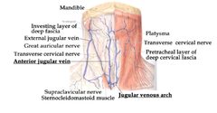
Superficial veins of the anterior cervical triangle

answer
There is venous return along the superficial aspect of the anterior cervical region The external jugular vein is often times palpated and measured to determine if there is high venous pressure, i.e. CHF There is a jugular venous arch ?Bilateral anterior jugular veins drain the mandible and feed into arch ?Size of these veins is usually inversely proportional to the size o the external jugular vein ?If external jugular vein is very large, the anterior jugular and arch will be small, vice versa
question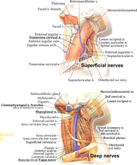
Nerves of the anterior cervical region

answer
Superficial nerves ?Transverse cervical nerve originates from cervical plexus in lateral cervical region, crosses over top of sternocleidomastoid muscle and supplies cutaneous innervation to neck Deep nerves ?Glossopharyngeal nerve branches ?Hypoglossal nerve ?Both sets of nerve branches innervate the tongue
question
Root of the neck
answer
Just above the clavicle and is defined as structures that are anchoring neck to thorax Structures that pass up through the root of the neck and pass down through here are: ?Esophagus beginning in neck and passing down to thorax ?Trachea, from oral cavity into thoracic region ?Internal carotid artery and subclavian artery exit from thorax via arch of aorta and enter into neck ?Internal jugular vein and subclavian vein come through neck and enter thorax ?Phrenic nerve is coming out of the cervical plexus through the root of the neck, passing through thorax to innervate diaphragm ?Trauma can cause loss of diaphragm function and patient must be placed on ventilator ?Vagus nerve (CN X) parasympathetic innervation to thorax and abdomen ?Found in carotid sheath ?Travels deep to internal jugular vein and carotid artery ?At risk when you do surgery on carotid artery ?Recurrent laryngeal nerve is a motor branch of the vagus nerve ???Damage here can impact vocalization ???Path of nerve is different from right to left ?Sympathetic trunk ?Continuous w/ sympathetic trunk that is in thorax ?There are cervical ganglia along trunk, largest is superior cervical ganglion, which is very deep ?Also has middle and cervicothoracic ganglion ?Thoracic duct leaves root of neck and travels to head region ?Branches of subclavian artery ?Internal thoracic ?Vertebral ?Costocervical trunk Arteries that are at risk from trauma to this region are branches from subclavian artery, branches from brachiocephalic vein, internal jugular and subclavian veins
question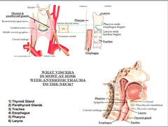
Viscera of the neck

answer
The most superficial structure of the viscera is the thyroid gland ?sits anterior to the trachea ?isthmus is sitting below C6 and the lobes travel superiorly The pharynx blends with the esophagus at the level of C6 ?With anterior trauma the larynx and trachea are the most at risk ?C6 is a good barometer for the division between larynx, pharynx, trachea and esophagus. ?With trauma to the neck you have to think that the structures that are most at risk are the larynx and trachea Thyroid gland ?Bilobed gland that is responsible for metabolism. ?Highly vascularized with superior and inferior branches traveling to it ?The superior blood supply comes from a branch of the external carotid ?The inferior thyroid artery is a branch from the thyrocervical trunk from the subclavian artery ?Large venous distribution because we want the signals that they thyroid is producing to be put into circulation quickly ?There is the superior, middle and inferior thyroid veins ?Thyroid ima artery sometimes comes directly off the subclavian and supplies the isthmus to the thyroid ?There is usually not that much blood supply to the midline of the neck, but in tracheotomy or other procedures that revolve around cutting at the midline, you have the potential to cut the thyroid ima artery and cause bleeding. ?If you take out the thyroid gland the recurrent laryngeal nerve is at risk and you can impact vocalization ?runs directly behind the lobes of the thyroid through the cricothryoid membrane and into the larynx to supply motor innervation to the muscles of the larynx. ?Enlarged thyroid (goiter) does not typically compress posterior structures because it expands outward. ?To check if the thyroid is functional you give radioisotope of iodine and look for hot and cold regions through a scintigram ?Hot region means that it is functional ?Cold region means that the thyroid is non-functional and not taking up the iodine. Parathyroid glands ?Lie on the posterior aspect of the thyroid ?Four total glands, two on the superior portion and two on the inferior portion of the thyroid. ?Regulate calcium and phosphorus in the blood. Trachea Esophagus Pharynx Larynx
question
Pharyngeal pouches and arches
answer
Humans have 5 arches (1, 2, 3, 4 and 6) ?Never see more than 4 at a time All grooves are lined by ectoderm ?Pouches are lined by endoderm ?No continuity ? double layer membrane b/w every arch Inside each arch, there are 4 major things that develop w/in each arch ?Major blood vessel (aortic arch) ?Cranial nerve ?A piece of musculature (from occipital somites) ?Piece of cartilage Normally, tissue in each arch is mesoderm ?However, cartilage will have small contribution from neural crest Pouches are b/w the arches
question
Nerve Development in Pharyngeal Arches
answer
1st arch gets maxillary and mandibular branches of the trigeminal nerve ?all muscles of mastication get mandibular branch 2nd arch will get CN VII (facial nerve) ?both motor and sensory component 3rd arch - CN IX (glossopharyngeal) 4th and 6th arch each get different parts of vagus ?4th - superior laryngeal nerve ?6th - recurrent laryngeal nerve Facial nerve (CN VII) will send a branch to the 1st arch ? chorda tympani CN IX will send a branch to the 2nd arch
question
Muscles of Pharyngeal Arches
answer
Majority of muscles from 1st arch are muscles of mastication (Masseter, temporalis, medial and lateral pterygoids) ?Also muscles of floor of mandible, which get mandibular branch (mylohyoid, anterio belly of the digastric, tensor vali palatine, tensor tymani muscles) Muscles from 2nd arch are muscles of facial expression ?Include auricularis, buccinators, platsma frontalis, orbicularis oris and oculi ?Also includes posterior belly of digastric, stapedius, and stylohyoids The only muscle of the third arch is the stylopharyngeus muscle Arches 4 and 6 are involved w/ pharyngeal muscles and laryngeal muscles ?4 ? mostly pharyngeal muscles but also one laryngeal muscle (cricothyroid) ?6 ? laryngeal muscles, all within larynx itself (cricoarytenoid, arytenoid, thyroarytenoid, vocalis) ?get recurrent laryngeal nerve
question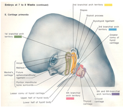
Cartilage of Pharyngeal Arches

answer
Cartilage can do several different things ?can remain separate ?can fuse to make one cartilage segment ?can form ligaments ?can ossify to form bone 2 bones of middle ear come from cartilage of 1st arch, ossify to form bone ?middle part regresses to form 2 ligaments ? sphenomandibular ligament, and the small anterior ligament of the malleus ?ventral portion is a special kind of cartilage (Meckel's) that is replaced by the mandible Dorsally, 2nd arch forms the 3rd bone of middle ear (stapes), styloid process of temporal bone ?Intermediate portion forms the stylohyoid ligament ?Ventral portion ossifies to form lesser cornu of the hyoid bone, upper body of hyoid bone 3rd arch ossifies to form greater cornu of hyoid, lower body of hyoid bone 4th arch - forms laryngeal cartilage ? thyroid, arytenoid, comiculate, cuneiform 6th arch forms cricoid cartilage
question
Blood Vessels of the Pharyngeal Arches
answer
1st arch forms parts of maxillary and external carotid arteries 2nd arch forms stapedial artery ?most degenerates 3rd arch ?proximal part forms common carotid artery ?distal part forms internal carotid artery 4th arch ?left: part of arch of aorta ?right: proximal part of subclavian artery 6th arch ?Left: proximally orms left pulmonary artery, distally forms ductus arteriosus ?Right: proximally forms right pulmonary artery, distally degenerates
question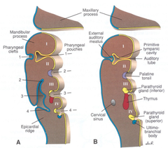
Pharyngeal pouches

answer
Pouch 1 becomes the tubotympanic recess, which forms the tympanic cavity and auditory tube Pouch 2 gives rise to palatine tonsils ?They will stay roughly where they are forming Pouch 3 ?Dorsal bud gives rise to inferior parathyroids ?Ventral bud gives rise to the thymus gland, which migrates into 4th intercostal space ?Derivatives migrate away, more so than derivatives of 4th pouch Pouch 4 ?Dorsal bud gives rise to superior parathyroids ?Ventral bud gives rise to ultimo-branchial body, the C-cells of the thyroid gland ?Migrates to thyroid gland Pouch 5 does not form
question
Branchial Sinus, Cyst or Fistula
answer
Occurs when a pharyngeal groove or pouch persists (most often 2nd groove/pouch) If persistent groove remains open onto skin or the persistent pouch remains open into the pharynx, a branchial sinus results If the persistent groove or pouch closes over at the surface so that there is no communication w/ skin or pharynx, a branchial cyst results ?Cyst may become infected and secondarily break through to the surface ?A persistent cervical sinus may also give rise to branchial cyst When the same groove and pouch persist (most often 2nd groove and pouch) on the same side, the pharyngeal membrane b/w them breaks down, leaving a patent duct-like branchial fistula which usually passes b/w external and internal carotid arteries and opens onto the skin at one end and into the pharynx at the other Sinuses and fistula most often open into the pharynx at the palatine tonsillar fossa and onto the skin in the lower 1/3 of the neck along the anterior border of the sternocleidomastoid muscle Mucus may be discharged from sinus and fistulas Uncommon, originate in 6th-7th weeks, appears during infancy
question
Congenital Thymic Aplasia and Absence of Parathyroids Glands (DiGeorge's Syndrome)
answer
Results when the 3rd and 4th pharyngeal pouches fail to give rise to the thymus and parathyroid glands This condition leads to congenital hypoparathyroidism and infection and is usually fatal within a few months after birth Thymic aplasia may also occur alone Infant usually succumbs to infection Very rare, originate 5th-6th week, appears at birth
question
Cervical thymus & accessory thymic tissue
answer
Results when the thymus or part of it fails to descend into the anterior and superior mediastinum from the third pharyngeal pouch Most often a cord of thymic tissue remains behind in migration path of the gland Very rare, origin 8th-9th week, appears during infancy
question
Ectopic and Variations in Number of Parathyroid Glands
answer
Ectopic parathyroid glands result either b/c the glands fail to migrate or b/c they migrate too far (w/ thymus into mediastinum) ?May lodge anywhere along migratory path Supernumerary glands result from division of one or more parathyroid gland primordia and hypoparathyroidism from the failure of one or more primordia to develop ?50% of population has an abnormal number of parathyroid glands ?most common number of abnormal parathyroid glands is 2 or 3 although up to 8 have been recorded Common, origin 6th-7th week, appears at birth
question
Thyroid Gland
answer
Origin: 4th week as an endodermal diverticulum in mid floor of pharynx at the level of the 2nd arch Descends into neck but maintains its connection to pharynx via the thyroglossal duct and opens onto the tongue through the foramen cecum ?Thyroglossal duct normally disappears by 7 weeks of development ?In 50% of population the lower part of the thyroglossal duct remains as a pyramidal lobe of the thyroid gland
question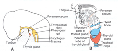
Thyroglossal Ducts & Sinuses

answer
Thyroglossal duct is formed as thyroid gland descends and it connects the gland to the foramen cecum of the tongue The inferior end of the duct persists as the pyramidal lobe in 50% of the population, the rest usually degenerates If the superior part fails to atrophy, cysts may form ?Leaves bulges in the anterior midline of the neck if these cysts become infected they may rupture the overlying skin and form a sinus (occurs in 1/3 of all thyroglossal duct cysts) open permanently to the surface ?Sinuses open most often in the midline of the neck anterior to the laryngeal cartilage
question
Tongue Development
answer
Origin: begins during the 4th week of development Develops in different parts: anterior 2/3 and poster 1/3 Development of the anterior 2/3 of tongue ?From 2 lateral (distal) tongue buds (lateral lingual swellings) arising in the mesoderom of the floor of the pharynx opposite the first pair of pharyngeal arches ?The two lateral tongue buds fuse in the midline represented by the median sulcus and septum of the tongue ?Median tongue bud (tuberculum impar) does form but it is quickly overgrown by the 2 lateral tongue buds and disappears ? no adult structures derived from median tongue bud Development of the posterior 1/3 of tongue ?The hypobranchial eminence, a mesodermal swelling in the floor of the pharynx opposite pharyngeal arches III and IV, gives rise to the posterior 1/3 of tongue and to the epiglottis ?Small structure called the copula does form in the floor of the pharynx opposite the second pair of pharyngeal arches but it is overgrown by hypobranchial eminence and disappears ? no adult structures from copula Fusion of anterior and posterior parts of tongue is demarcated by a v-shaped line called the terminal sulcus Musculature is derived from the occipital myotomes which migrate into the tongue Innervation ?Motor: CN XII (hypoglossal) ?Sensory of anterior 2/3 of tongue ?lingual branch of mandibular nerve (V) for touch, temp, etc. from pharyngeal arch I ?chorda tympani (from facial) to fungiform papillae for taste from pharyngeal arch II ?Sensory of posterior 1/3 of tongue ?Glossopharyngeal nerve (IX) from pharyngeal arch III to vallate taste buds and mucosa ?Vagus nerve (X) from pharyngeal arch IV to base of epiglottis
question
Cleft or Bifid Tongue
answer
Results when lateral tongue buds (lateral lingual swellings) fail to fuse properly in the midline If fusion is incomplete, a cleft tongue results in the posterior part (over tuberculum impar) of the anterior tongue If the swellings remain completely separate, a bifid tongue results Rare, origin 4th-5th week, appears at birth
question
Vestibulocochlear Organs
answer
?Internal Ear ?Middle Ear ?External Ear
question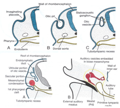
Development of Internal Ear

answer
Origin: early 4th week ?First of three ear divisions to form Development ?Otic Placodes arise as ectodermal thickenings on each side of developing hindbrain ?Placodes invaginate to form otic pits and soon lose their connection with the overlying ectoderm ?These otocysts (vesicles) are the primorida of the membranous labyrinth, they different into two regions ???Dorsal region - utricular portion ???Ventral region - saccular portion ?Utriculus gives rise to: ?Endolymphatic duct ?Semicircular ducts ?Cristae ampullares and maculae utriculi of the semicircular ducts and utricle respectively and they contain sensory receptors ?Saccule gives rise to: ?Maculus sacculi of the saccule which contain sensory receptors ?Cochlea - arises as tubular diverticulum from saccule, duct coils as it grows ?Organ of Corti - differentiates from ectodermally derived lining of the cochlear duct, innervated by fibers from cochlear ganglion of CN VIII ???Ganglionic cells of CN VIII migrates along the cochlea to form the spiral (cochlear) ganglion which innervate the organ of Corti ?Otic Capsule ?The mesenchyme surrounding the otocyst differentiates into a cartilaginous otic capsule ?Otic capsule ossifies to form the bony osseous labyrinth of the inner ear (includes the semicircular canals) ?In addition the optic capsule forms the perilymphatic space around the membranous labyrinth - space differentiates further into scala tympani and scala vestibule
question
Development of Middle Ear
answer
Origin: begins during late 4th week, early 5th week Development ?Derived from 1st pharyngeal pouch ?Pouch elongates into a tubotympanic recess which envelopes the bones of the middle ear ?The distal portion of the recess expands to become a tympanic cavi and it is the endodermal lining of this cavity which will envelop the bones of the middle ear (malleus, incus, stapes), their ligaments and tendons and the chorda tympani nerve (from CN VII) ?The tympanic cavity also gives rise to the tympanic (mastoid) antrum during late fetal life, which communicates w/ mastoid air cells
question
Development of External Ear
answer
Origin: begins during 5th week (external auditory meatus) and 6th week (auricle) Development ?External acoustic meatus - ectodermal lining of the 1st pharyngeal groove begins to proliferate, occludes the groove and extends inwards as a solid mass of ectodermal cells ?Mass later hollows out during late fetal life by degeneration of the ectodermal cells in the center of the mass ?Tympanic membrane - as the first groove and pouch come together they are initially separated only by a bilaminar, membrane of ectoderm and endoderm ?Mesoderm soon moves in b/w these two epithelial layers to give rise to fibrous element of tympanic membrane ?Auricle - arises from 6 mesodermal swellings (auricular hillocks) of the first and second pharyngeal arches around the margins of the first pharyngeal groove ?The auricles begin forming in the upper neck region but ascend to the level of the eyes on the sides of the head as the mandible forms ?Nerve supply ?Arch 1 derivatives are innervated by mandibular branch of trigeminal nerve ?Arch 2 derivatives are innervated by some cutaneous branches from facial (CN VII) nerve near the mastoid process but most of arch II derivatives receive cutaneous innervation from the cervical plexus (lesser occipital, greater auricular nerves)
question
Congenital Abnormalities of the Ear
answer
Since the middle and inner ear develop independently, impaired hearing can be due to either a perceptive deafness (neurosensory loss in the inner ear) or a conduction deafness (inability to conduct sound across middle ear)
question
Inner Ear Abnormalities
answer
Perceptive deafness Most types of congenital deafness are genetic in origin and 25% of those are caused by dominant gene Spiral Organ of Corti is absent and the spiral ganglion deficient in neurons so that sound cannot be perceive by nerves of inner ear Rubella - causes congenital deafness since it destroys the epithelail lining of the cochlea and prevents the formation of the Organ of Corti ?Critical period for infection is during the 1st trimester but the damage incurred may not be detected until 1-4 years postnatally
question
Middle Ear Abnormalities
answer
Conduction deafness Conduction deafness most often involve defects in the formation of the bones of the middle ear but it can also occur if an ectodermal epithelial plug persists in the external meatus or if the tympatic membrane is damaged Sound is prevented from reaching spiral organ of corti in inner ear
question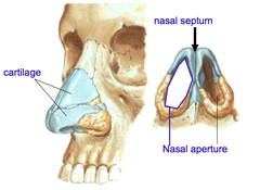
Nasal Skeleton

answer
On the distal most tip of nose - cartilage Nasal aperatures allow for air to passage through ?are divided by nasal septum
question
5 Bones in Nasal Septum
answer
1. ethmoid (perpendicular plate) 2. sphenoid 3. vomer - contributes to septum 4. palatine - primarily contributes back portion of hard palate 5.maxilla
question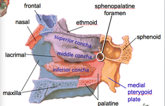
Lateral Wall of Nasopharynx

answer
Can no longer see the vomer (which really only contributes to septum) Concha ?Superior, middle or inferior ?Has meatus inferior to each
question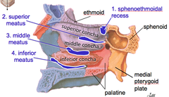
4 Spaces of Lateral Wall of Nasopharynx

answer
1. sphenoethmoidal recess 2. Superior meatus (inferior to superior concha) 3. Middle meatus (inferior to middle concha) 4. Inferior meatus (inferior to inferior concha)
question
4 Paranasal Sinuses Drain into Meatuses
answer
parallel to midline Frontal sinus ?Most superior Sphenoid sinus ?In sphenoid bone itself Ethmoid Sinus ?On lateral aspect of ethmoid bones, just past cribriform plate Maxillary Sinus ?Within maxilla ?Roots of teeth approach this sinus
question
Lacrimal gland and nasolacrimal duct
answer
If you are producing tears, the lacrimal gland up in the upper lateral portion of orbit will produce them, which will go in and around eyeball ?Will eventually be drained into lacrimal duct, which opens up into the inferior meatus
question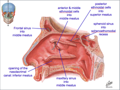
Sinuses Connecting To Meatuses

answer
Lacrimal duct opens up into inferior meatus ?Frontal sinus opens up into the middle meatus ?Anterior aspect and middle aspect of ethmoidal cells also open into middle meatus ?Maxillary sinus ALSO drains into middle meatus Posterior ethmoidal cells open into superior meatus Sphenoid sinus drains into the spenoethomoidal recess THESE TEND TO WORK BEST WHEN YOU ARE UPRIGHT ?When you lay down, things can get blocked and it can cause congestion
question
Blood Supply and Somatic Sensation
answer
Major contributors are essentially associated with ethmoidal branches (from internal carotid) and greater palatine branches (also from internal carotid) ?superior labial artery (superficially) is a branch from facial artery, from external carotid) Kiesselbach's plexus - area that will break first if you get punched in the nose b/c it is cartilage ?epitaxis = nose bleed ?maxillary division of trigeminal responsible for pain here
question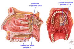
Palatine Nerves

answer
Greater palatine nerve takes care of hard palate Lesser palatine nerve takes care of soft palate ?Goes in posterior direction towards soft palate
question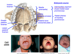
Palatine Foramina

answer
Greater palatine foramen has larger nerve/vascular bundles, is more anterior than lesser palatine ?Located in inferior portion of hard palate Cleft palate - didn't properly fuse in one way or another ?Intermaxillary segment is most anterior ?Palatal shelves of maxillary prominences is more posterior
question
Pharyngeal Arch 1

answer
Aka mandibular arch Skeletal Derivatives ?Malleus ?Incus ?Sphenomandibular ligament ?Meckel's cartilage (future mandible) Muscular Derivatives ?Mm. mastication ?Anterior belly of digastric m. ?Mylohyoid m. - goes to hyoid bone ?Tensor tympani m. ?Tensor veli palatini m. - inserts of lateral aspect of soft palate, tenses soft palate and widens it out Innervated by CN V3
question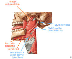
Pharyngeal Arch 2

answer
Aka hyoid arch Skeletal Derivatives ?Stapes ?Styloid process ?Stylohyoid ligament ?Lesser horn ; upper body of hyoid bone Muscular Derivatives ?Stapedius m. ?Mm. facial expression ?Posterior belly of digastric m. ?Stylohyoid m. Innervated by Facial n., CN VII
question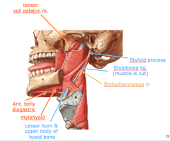
Pharyngeal Arch 3

answer
Skeletal derivatives ?Greater horn and lower body of hyoid bone Muscular Derivatives ?Stylopharyngeus msucle Innervated by CN IX
question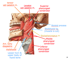
Pharyngeal Arches 4 ; 6

answer
Laryngeal cartilages: ?Epiglottis ?Thyroid cartilage ?Cricoid cartilage ?Arytenoid cartilage ?Corniculate cartilage ?Cuneiform cartilage Muscular derivatives and innervation ?All palatine muscles. (CN X) ?Except tensor veli palatine muscle - CN V3 ?All pharyngeal muscles. (CN X) ?Except stylopharyngeus muscle - CN IX ?All laryngeal muscles (CN X) ?Cricothyroid muscle - superior laryngeal nerve ?Intrinsic muscles - recurrent laryngeal nerve
question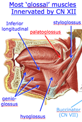
Most glossal muscles innervated by CN XII

answer
Innervated by hypoglossal nerve (CN XII) EXCEPT palatoglossus ? innervated by vagus nerve
question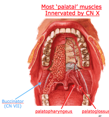
Most palatal muscles innervated by CN X

answer
Vagus nerve EXCEPT buccinator, which is innervated by CN VII (facial)
question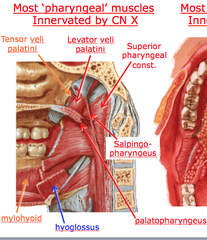
Most pharyngeal muscles innervated by CN X

answer
Salpingopharyngeus merges with palatopharyngeus ?Superior pharyngeal constrictor Hyoglossus is innervated by hypoglossal CN XII
question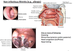
Non-Infectious Rhinitis

answer
Allergies ? congestion, sniffles, sneezing, runny nose, irritation w/ mucosal lining w/ allergic reaction, concha can swell, which can impinge on ability to breath well doesn't necessarily mean there will be pain ? just irritation
question
Toothache
answer
Dental pain can often refer into facial pain V2 has branches associated with teeth, as well as somatic sensory to skin of midface, nasal cavity, maxillary sinuses and palate
question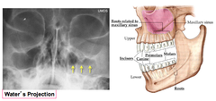
Maxillary Sinusitis

answer
Cloudiness in X-ray in maxillary sinus is indicative of liquid forming in there ?Drains into middle meatus, comes out nose If tooth removed, opening can allow infection to pass up into space of maxillary sinus
question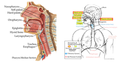
Resonance and Articulation

answer
Allows communication ?Humming and speaking/singing Nasal cavity, oral cavity and pharynx ? areas where sound can resonate, amplify Lips, tongue, vocal folds in larynx ? move in specific pattern to allow annunciation Sinuses can play a role in speaking as well ?Opening of sinuses has to be completely open to airway in order for them to have an effect ?If you hum, and then you open move and work to keep tongue down ? connectivity lost b/c airway blocked b/c tongue and soft palate were pushed near each other Both talking and humming are affected by nasal airways ? i.e., voice different when you have a cold
question
Nasal Airflow
answer
Laminar - quiet inspiration High inspiratory rate, sniffing ? turbulent
question
Chemosensation
answer
Flavor is both taste (from taste buds) and smell (from olfactory epithelium) Additionally, somatosensory overtones play a role ?"trigeminal sense," (hot, cold, tingling, irritating) ?related to texture, etc.
question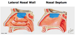
Olfactory Epithelium

answer
Olfactory epithelium is in the upper region of the nasal cavity ?This is why airflow is important ?Redirecting air with concha is helpful in making sure air gets to olfactory epithelium, facilitating smell Cribriform expands over it, w/ nerve endings piercing it and entering mucosa BLUE is where most of the sense of smell is
question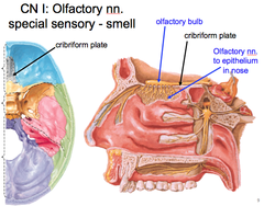
CN I: Olfactory Nerve

answer
Special sensory - smell Forms olfactory bulb, which pokes through cribriform plate, forming olfactory nerves to epithelium in the nose
question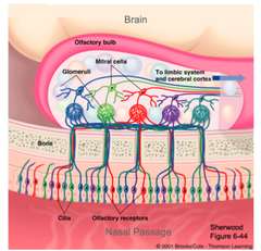
Olfactory receptor cells

answer
Olfactory receptor cells synapse w/ mitral cells in the olfactory bulb (olfactory glomeruli) Individual glomeruli respond selectively to distinct oderants
question
Trigeminal Sense
answer
Trigeminal nerve (CN V) provides most of the sensory innervation of the face: mechanical, thermal, proprioception, nociception Also sensitive to chemical irritants in nose, oral, cavity, eye Examples: ?Noxious - ammonia ?Cool - menthol, camphor ?Hot - spicy food ?Sharp - ethyl alcohol Means that food scents can serve as a warning factor, as well as can stimulate hunger
question
Mouth Watering
answer
Major salivary glands and ducts have protective and digestive functions ?Moisten oral mucosa ?Aid swallowing - moisten dry food ?Dissolve/suspend foods to stimulate taste buds ?Buffer oral cavity contents - high [bicarbonate ions] ?Digestion (carbohydrates via ?-amylase) ?Control bacterial flora of oral cavity (IgA, lysozyme) ?Protect teeth via saliva protein coat (antibacterial compounds) ?PARASYMPATHETIC - Abundant, watery saliva ?SYMPATHETICS - Scant, viscous saliva ?via external carotid plexus Parotid (all serous saliva) CN IX via otic ganglion Submandibular (mostly serous saliva) - CN VII via submandibular ganglion Sublingual (most mucous saliva) - CN VII via submandibular ganglion
question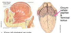
Tongue

answer
Has a core of skeletal muscle Wide range of muscles associated w/ moving tongue ?the stuff coming off the tongue = genioglossus ? looks like it has a few divisions but it doesn't, is the major muscle for sticking out the tongue ?can test for hypoglossal nerve deficits by having patient stick out tongue ?should stick out straight ?if there is a deficit, the tongue will push itself towards deficit ?i.e. left side deficit means tongue will extend towards the left cheek ?deep to that is the geniohyoid ? doesn't move tongue, only going from chin back towards hyoid Surface of tongue has a terminal surface that separates portions of the tongue ?Oral portion is in the mouth, more anterior ?Pharyngeal portion is the root, the part sticking in towards the throat Dorsum is covered w/ papillae ?Lamina propria folded up w/ stratified squamous epithelia (somes keratinized)
question
Entire tongue is sensitive to all tastes
answer
Bitter, salty, sweet, sour and umami are distributed evenly throughout tongue
question
Anterior vs. Posterior Tongue

answer
Anterior 2/3 of tongue (oral) ?Taste - facial nerve (Chorda tympani) ?Touch - trigeminal nerve (lingual nerve) Posterior 1/3 (pharyngeal) ?Taste and touch - glossopharyngeal nerve Gag reflex ?Afferent limb: glossopharyngeal nerve Also, some extrapapillary taste buds scattered in palate and pharynx
question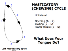
Masticatory (Chewing) Cycle

answer
Unilateral ?Opening (6-2) ?Closing (2-5) ?Power Stroke (5-6) What does you tongue do? ?Moves the whole time, to move food to right shape/side, gets it ready to swallow
question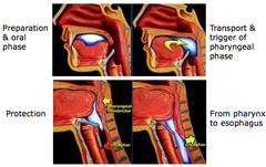
Swallowing Reflex

answer
Preparation and oral phase ? transport and trigger of pharyngeal phase ? protection ? from pharynx to esophagus Want to make sure you can get the food back and down esophagus without getting into nasal cavities or trachea
question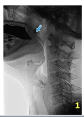
Swallowing Reflex - Preparatory Phase

answer
Base of tongue fauces & soft palate close oral cavity posteriorly Prevents spillage of food into pharynx (nasal region) and open larynx
question
Swallowing Reflex - Oral Phase
answer
Propels bolus from oral cavity into pharynx Base of tongue, hyoid and larynx, and pharynx move craniad Soft palate elevates to prevent spillage into nasopharynx Larynx closes (contraction of aryepilgottic folds) Dorsum of posterior tongue depresses and fauces relax ? move bolus into oropharynx
question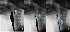
Swallowing Reflex - Pharyngeal Phase

answer
Bolus transport from oropharynx into esophagus w/o aspiration Contraction of upper pharyngeal constrictor Contraction of middle pharyngeal constrictor Contraction of lower pharyngeal constrictor, relaxation of cricopharyngeal muscle
question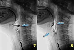
Post-swallowing/Repositioning

answer
Epiglottis elevates to regain its resting position Larynx opens
question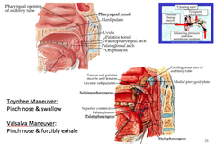
Connectivity to Middle Ear

answer
As you are swallowing/yawning, you are adjusting aperture associated with auditory tube ? can change pressure associated with middle ear ?Have tympanic membrane that blocks it from outer ear, so this pressure changes happens on the inside Children who have their tonsils out can have infections of this area, and inadvertently give themselves inner ear infections ?Cleft palate patients are more prone to having this happen b/c palatal muscles are not where they are supposed to be
question
Esophageal Dysphagia
answer
Characterized by difficulty swallowing several seconds after initiating a swallow Could be obstructive (difficulty w/ solids) Could be motor loss (difficulty w/ solids and liquids) similar to patients with oropharyngeal dysphagia, dysphagia related to distal esophageal disease, such as a peptic stricture, may sometimes be felt in the suprasternal notch ?However, patients with oropharyngeal dysphagia have difficulty initiating a swallow and may have nasopharyngeal regurgitation, aspiration, and a sensation of residual food remaining in the pharynx ?In contrast, esophageal dysphagia is characterized by difficulty swallowing several seconds after initiating a swallow
question
Globus Sensation
answer
Globus sensation is defined as a persistent or intermittent nonpainful sensation of a lump or foreign body in the throat with the occurrence of the sensation between meals and the absence of dysphagia, odynophagia, an esophageal motility disorder, or gastroesophageal reflux as the cause of symptoms ?These criteria must be fulfilled for the last three months with symptom onset at least six months before a diagnosis of globus sensation can be made Pts w/ globus sensation (sensation of lump in throat) can sometimes be confused as oropharyngeal dysphagia ?In contrast with patients with oropharyngeal dysphagia whose symptoms occur with swallowing, patients with a globus sensation report the sensation of a lump or foreign body independent of swallowing
question
Oropharyngeal Dysphagia

answer
Diagnosis of oropharyngeal dysphagia is based on a history of difficulty transferring food from the mouth to the pharynx and report a feeling of an obstruction in the neck Other common complaints include coughing, choking, drooling, and regurgitation when swallowing liquids or solid food Patients may have a history of aspiration pneumonia and weight loss. Oropharyngeal dysphagia is an alarm symptom that warrants urgent evaluation ?It is important to determine the underlying etiology and to assess the severity of oropharyngeal dysfunction prior to initiating treatment ?A careful history can distinguish between an esophageal and oropharyngeal cause of dysphagia and globus sensation and may help identify the underlying etiology
question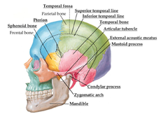
Bone Of Intratemporal Fossa

answer
2 temporal lines ?run though frontal and parietal bones as well pterion - 4 bones contribute (frontal, parietal, sphenoid, temporal bone temporal bone and zygomatic bone contribute to zygomatic arch ?part of temporal bone that contributes is the zygomatic process ?part of the zygomatic arch has the condylar process from the mandible coming off of sphenoid bone, we have processes that extend inferiorly ?medial and lateral pterygoid plate
question
Mandible
answer
Mandibular condyle (condylar process) is the part that articulates w/ TMJ, is encapsulated ?Has a head and a neck Mandibular notch is b/w coronoid process and the head/neck of condylar process Has mental foramen ?Trigeminal nerve V3 (mandibular) exits here as the mental nerve Mandibular foramen has inferior alveolar nerve enter it ?Provides sensory fibers to teeth ?Root canals basically obliterate these nerve branches Mylohyoid groove has mandibular branch of trigeminal ?This nerve provides inneration to mylohyoid and the digastric Genio tubercle - tubercle on anterior aspect of mandible Sublingual fossa is right in the midline Angle of the mandible (gonion) Just anterior to mandibular foramen is the lingula, a tongue like process ?Sphenomandibular ligament attaches here, important for stabilization of the jaw as it moves
question
Temporal Region
answer
Posterior - Superior temporal line Anterior - Frontal and zygomatic bones Lateral - Zygomatic arch Inferior - Infratemporal crest Floor - Pterion Roof - Temporal fascia
question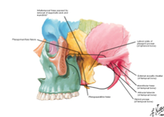
Infratemporal Fossa

answer
Posterior - tympanic plate of the mastoid and styloid processes of the temporal bone Anterior - Posterior aspect of maxilla Lateral - Ramus mandible Medial - Lateral pterygoid plate Superior - inferior surface of the greater wing of the sphenoid Inferior - attachment of medial pterygoid to mandible near its angle
question
Content of Infratemporal Fossa
answer
Muscles ?Temporalis, lateral and medial pterygoid ?Masseter Blood Vessels ?Maxillary artery ?Pterygoid venous plexus Nerves ?Mandibular division of trigeminal ?Chordatypani ?Otic ganglion
question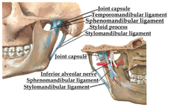
Temporamandibular Joint

answer
Mainly movement involved in mastication, movement to facilitate speech Synovial joint ? has a capsule ?Capsule covers condylar region and extends down to neck of mandible Stabilizing the joint are 2 ligaments ?Stylomandibular ligament is attached to the styloid process - limits jaw from moving too far anteriorly ?Main stabilizing joint is the sphenomandibular ligament, which is attached to sphenoid bone and lingual ? limits jaw from going too far anteriorly AND laterally during chewing Mandibular fossa - when condyle of mandible articules ?is articular cartilage b/w bones, which must move as jaw opens ?Can get arthritis here, like any other joint ?Best theory for clicking sound in jaw: articular cartilage is not moving appropriately, the clicking is the movement of the bone against the bone
question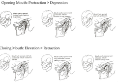
Movement of the TMJ

answer
Elevation = closing the mouth ?temporalis, masseter, and medial pterygoid Depression = opening the mouth ?For the most part, due to gravity ?If we have one muscle that generates force, it is the lateral pterygoid Protrusion = Protrude the chin ?Lateral pterygoid (prime mover) masseter and medial pterygoid Retrusion = retrude the chin ?Temporalis and masseter Lateral movement = grinding and chewing ?temporalis of same side, pterygoids of opposite side and masseter. When we open our mouth, we get a little forward movement (protusion) and THEN we get swinging hinge movement ?When we close our mouth, we elevate then retrude mandible back to resting position in TMJ
question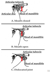
Dislocated TMJ

answer
TMJ can dislocate Excessive depression and protrusion can sometimes cause an anterior dislocation of the joint The tone of the muscles in this area means that the joint will not slip back into place ?Individual will be in pain, will not be able to close their mouth ?Usually just takes physical manipulation of condyles to reset joint Anterior tubercle prevents anterior dislocation in normal movement ?When you do get dislocation, this structure becomes problematic and does not allow the joint to easily slip back into place
question
Muscles of Mastication
answer
Temporalis Masseter Lateral pterygoid Medial pterygoid Assisting in mastication ?Infrahyoid and suprahyoid ?Platysma
question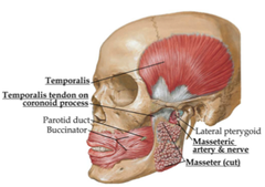
Temporalis

answer
Attachments: Sits in temporal fossa; attaches to coronoid process and anterior border of ramus of mandible Innervation: Mandibular division of Trigeminal Actions: Elevates mandible (main function), retraction and lateral movement ?Posterior fibers that run almost horizontally are what help retract ?Working unilaterally, it would cause lateral movement
question
Masseter
answer
Attachments: zygomatic arch; Angle and lateral surface of ramus of mandible Innervation: Mandibular division of trigeminal Action: Elevates mandible, protrusion ?Mainly more anterior fibers would help to pull posterior part of mandible forward for protusion Fractures of the mandible can be damaging, and masseter provides some protection ?Weakest point of mandible is superior to where masseter attaches
question
Lateral pterygoid
answer
Attachment: Sphenoid bone and lateral side of lateral pterygoid plate; TMJ joint capsule and condyloid process of mandible Innervation: Mandibular division of trigeminal Action: Bilaterally protracts mandible and depresses chin; unilaterally swings jaw towards contralateral side (work back and forth for lateral chewing/grinding)
question
Medial pterygoid

answer
Attachments: Medial surface of lateral pterygoid plate and tuberosity of maxilla; medial surface of ramus to mandible inferior to mandibular foramen. (mirror image if ipsilateral masseter) Innervation: Mandibular division of trigeminal Action: Elevate mandible, protrusion, alternating unilaterally creates grinding
question
Maxillary Artery
answer
Branch from external carotid artery Pneumonic for branches: DAM I AM Piss Drunk, But Stupid Drunk I Prefer, Must Phone Alcoholics Anonymous Can be divided into 3 divisions based on relation to First part: DAM I A ?Deep auricular - External acoustic meatus; external tympanic membrane ?Anterior tympanic - internal tympanic membrane ?**Middle meningeal - Middle cranial fossa; trigeminal ganglion, geniculate ganglion, tensor tympani muscle ?**Inferior alveolar ?Accessory meningeal Second Part: M (from DAM I AM) ?**Muscular branches ?Masseteric ?Pterygoid ?Deep temporal ?buccal Third Part (most medial): Piss Drunk But Stupid Drunk I Prefer, Must Phone Alcoholics Anonymous (pterygomaxillary fissure) ?**Posterior superior alveolar ?Descending palatine artery ?Buccal (2nd part) ?**Sphenopalatine artery ?Descending palantine ?**Infraorbital ?Posterior superior alveolar ?Middle superior alveolar ?Pharyngeal branch ?Anterior superior alveolar ?Artery of pterygoid canal
question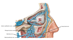
Pterygoid Venous Plexus

answer
Veins drain here from orbit, deep facial veins Blood can flow here to the re Damage to this area w/ slow continuous bleeding suggests involved of pterygoid venous plexus
question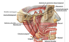
Nerves of Infratemporal Fossa

answer
Mandibular (most nerves here) ?Come through foramen ovale ?Lingular (sensory to anterior 2/3 of tongue) ?Chorda tympani (taste component for anterior 2/3, derived from facial nerve ?Also has parasympathetic to the submandibular gland ?Inferior alveolar (enters mandibular foramen, innervating alveoli of lower jaw, provides sensory innervation to parts of mandible) ?Gives off nerve to mylohyoid ?Auriculotemporal (sensory to area anterior to ear, over parotid gland) ?Sensory function isn't very important b/c a lot of nerves overlap here, providing sensory information ?Highlighted clinically b/c of otic ganglion Otic ganglion - ganglion for glossopharyngeal nerve (CN XI does tactile and taste for posterior 1/3 of tongue ?Preganglionic nerves from CNIX, hitches right w/ auriculotemporal ?Postganglionic nerves innervate parotid gland
question
Laryngeal Cartilage
answer
Thyroid ?Hyaline cartilage ?Open in the back, w/ attachments for membranes that run over it ?Has prominence (larger in men due to greater presence of androgens) ?The larger this is, the deeper the voice cricoid ?ring, where posterior surface has broad flat surface ?first complete ring you find in the airway ?limits the size of what will fit down the airway arytenoid (2) ?pyramidal hyaline cartilage ?structures that are moving as we move our true vocal cord ?have muscular surface where muscles attach to move them ?anterior portion is vocal part, w/ ligaments that run anteriorly ?can abduct, adduct, rotate laterally/medially, can rock back and forth anteriorly/posteriorly ?helps us to create pitch and inflection epiglottis ?elastic cartilage ?sits at back of tongue, prevents food from getting into laryngeal inlet corniculate (2) ?sit on top of arytenoid cartilages cuneiform (2) ?w/in arytenoid cartilages
question
Ligaments and Membranes of the Larynx
answer
**Thyrohyoid Ligament ?Median thyrohyoid membrane ?Lateral thyrohyoid membrane (inferior part) - produces false vocal fold Conus Elasticus Cricothyroid Ligament Vocal ligament - attached to arytenoid cartilages and the back of the thyroid cartilage Thyroepiglottic ligament hyoepiglottic ligament quadrangular membrane - runs around back, closes off laryngeal inlet (where we get extension of lateral thyrohyoid membrane that forms false vocal fold) ?aryepiglottic ligament ?vestibular ligament
question
Interior of the Larynx
answer
Vestibule Vestibular folds ?vestibular ligament ?quadrangular membrane Ventricle Vocal folds ?vocal ligament ?lateral cricothyroid ligament ?vocalis muscle ?thyroarytenoid muscle (main component in true vocal fold) Infraglottic cavity
question
Extrinsic Laryngeal Muscles
answer
Suprahyoid ?Mylohyoid ?Geniohyoid ?Stylohyoid ?Digastric Infrahyoid ?Sternohyoid ?Sternothyroid ?Thyrohyoid ?Omohyoid
question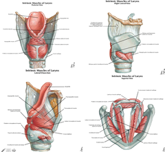
Intrinsic Laryngeal Muscles

answer
?Cricothyroid ?Lateral cricoarytenoid ?Posterior cricoarytenoid ?Transverse arytenoids ?Oblique arytenoids ?Thyroarytenoid ?Vocalis
question
Laryngeal Sphincters
answer
All intrinsic muscles except posterior cricoarytenoids are sphincter muscles ?Posterior cricoarytenoids - main abductor Think of them as adductors (move vocal folds medially) When they all work together, they close off the larynx
question
Abductors and Adductors of Laryngeal Folds
answer
Abductor ?Posterior cricoarytenoid Adductor ?Lateral cricoarytenoid (main adductor) ?Muscle fibers attach to back of arytenoid cartilages, runs posteriorly, pulls the back out laterally and the front medially
question
Tensor and Relaxer of Laryngeal Folds
answer
Tensor - allows for change of pitch, inflection ?cricothyroid - when it shortens, we increase the distance b/w arytenoid cartilages and the front of the thyroid cartilage, creating tension on vocal ligaments attached to arytenoid cartilage Relaxer ?Thyroarytenoid - shorten distance b/w arytenoid and thyroid cartilage
question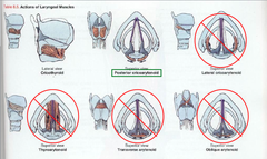
Innervation of Laryngeal Muscles

answer
Cricothyroid is innervated by external laryngeal nerve ?Damage can create result in inability to change pitch of voice ?Damage typically doesn't cause respiratory problems, unless it happens in a child whose respiratory muscles are not strong enough to compensate All other muscles are innervated by recurrent laryngeal nerve
question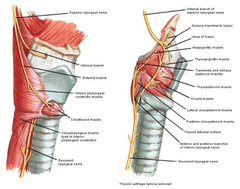
Nerves of the Larynx

answer
Vagus ?Superior laryngeal ?internal laryngeal ???Sensory above the vocal fold (senses particulate matter in upper airway, is part of cough reflex ?external laryngeal ???cricothyroid recurrent laryngeal ?inferior laryngeal
question
Arteries of the Larynx

answer
Superior laryngeal ?Branch of superior thyroid ?internal laryngeal nerve Inferior laryngeal ?Branch of inferior thyroid
question
Veins of the Larynx
answer
Superior laryngeal ?Drains into superior thyroid ?Drains into internal jugular vein Inferior laryngeal ?Drains into inferior thyroid ?Drain right into brachiocephalic vein
question
External Ear
answer
Ear (auricles) ?helix ?antihelix ?tragus ?antitragus External acoustic meatus ?lateral 1/3 cartilage ?medial 2/3 bone
question
Middle Ear
answer
roof (tegmen tympani) ?floor of the middle cranial fossa floor (jugular wall) ?bone between tympanic cavity and inferior jugular vein lateral (membranous) ?tympanic membrane medial (labyrinthine) ?inner ear anterior (carotid) ?bone between tympanic cavity and internal carotid artery posterior (mastoid) ?mastoid air cells
question
Tympanic Membrane
answer
Lateral border of middle ear Has embedded in it a tiny ossicle of the middle ear ?It creates ridges w/in tympanic membrane ?Want to see these, b/c it means that the middle ear does not have fluid accumulation Lateral process of the malleus runs through the ear ?Creates a small part of the pans tensa (tenser part of membrane) Umbo - most depressed part of membrane Anterior to umbo ? cone of light ?Will typically run anteriorly ?If you don't see it, it means the concavity of tympanic membrane is not there ? being push out by fluid
question
Ossicles
answer
Smallest bones in body ?Malleus ?Incus ?Stapes
question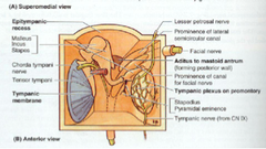
Malleus

answer
head ?articulates with incus lateral process handle ?embedded in tympanic membrane
question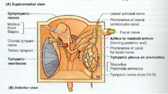
Incus

answer
Body Articulates with malleus Lenticular process articulates w/ stapes
question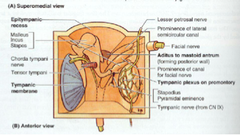
Stapes

answer
Head articulates with incus neck base articulates with oval window
question
Muscles of Middle Ear
answer
Assist in dampening sounds w/in inner ear Tensor tympani - stabilizes handle of malleus, limits amount of vibration tympanic membrane has, limits amount of vibration transmitted to stapes and incus ?Innervation by mandibular branch of trigeminal ?pharyngotympanic tube ?handle of malleus Stapedius (probably smallest skeletal muscle) - tense neck of stapes, trying to limit movement as it transmits to oval window ?posterior wall of tympanic cavity ?neck of stapes ?innervated by facial nerve(CN VII)
question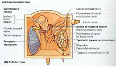
Nerves in Middle Ear

answer
Chorda tympani (branch of CN. VII) ?medial to neck of malleus ?superior to tensor tympani Tympanic nerve (branch of CN. IX) ?forms tympanic plexus on promontory of middle ear ?lesser petrosal nerve is a branch of this plexus ?penetrates roof of middle ear ?leaves skull through foramen ovale
question
Inner Ear
answer
Vestibulocochlear organ ?Bony labyrinth ?cochlea ?vestibule ???utricle ???saccule ?semicircular canals ?membranous labyrinth ?endolymph ?perilymph
question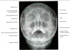
Evaluating Facial Bones w/ Radiology

answer
?Utilize frontal, lateral, and axial projection ?Look for symmetry ?Identify the midline ?Compare paired structures ?Notice different densities ?Look for calcifications ?Observe normal anatomic structures
question
Radiologic Evaluation of Facial Bones and Sinuses
answer
CT in coronal plane ? evaluate facial structures ?Excellent bony detail w/ 3D reconstruction MR - imaging study of choice to evaluate head and neck tumors ?Good soft tissue definition ?3D reconstruction ?in adults, lesion more likely malignant, which more likely benign in children
question
Sinusitis
answer
Symptoms ?Pain overlying sinuses ?Swelling ?Higher WBC count ?Want to check for local extension into surrounding soft tissues Early Changes ?Fluid collection (air/fluid level on erect film) ?Mucosal thickening Later changes ?Erosive or destructive changes involving wall of sinus ?Extension into cranium or the orbits
question
Thyroid
answer
Fusiform in shape Right and left lobes connected by an isthmus 5% have pyramidal lobe which is centrally located sometimes, internal jugular veins are asymmetric vertebral arteries usually asymmetric
question
Esophagus Muscles
answer
Inner muscles ? circular arrangement Outer muscles ? longitudinal arrangement
question
Pharynx
answer
Communicates w/ nasal and oral cavities Base of skull ; inferior border of cricoid cartiliage ; becomes esophagus 3 parts: ?Nasal: above soft palate ?Oral: b/w soft palate and larynx, continuous w/ mouth ?Laryngeal - posterior to the larynx
question
Larynx
answer
Originates at tip of epiglottis, extending inferiorly to inferior border of cricoid cartilage Hyoid bone IS NOT part of larynx
question
Facial Fractures
answer
Soft tissue injuries ?Extraocular muscle entrapment and impingement ?Superior, inferior, medial and lateral rectus muscles ?Orbital hematoma ?Globe rupture ?Dislocated lens ?Looks for foreign bodies ?Evaluate optic nerve Nasal fractures - most common Zygoma fractures - second most common
question
Tripod Fracture of Zygoma
answer
Lateral orbital wall of frontozygoma suture Maxillozygoma suture Zygomatic arch
question
Le Fort's Fracture
answer
facial fractures extend posteriorly towards pterygoid plate May result in facial bones being detached from cranium CT - visualize trapped soft tissue and nerves secondary to position of fracture fragments May require surgical decompression
question
Orbital Blowout Fracture
answer
Large object hits orbit Increased pressure in orbit causes fx of weakest walls Inferiorly - maxillary sinus (entrapment of inferior rectus) Medially - ethmoid sinus (via lamina papyracea)
question
Head and Neck Masses
answer
Adults ?Squamous cell carcinoma (most common malignancy) Children ?Benign ?Congenital cystic legions ???Thyroglossal cysts, branchial cleft cysts ?Teratoma ?Juvenile angiofibroma ?Malignant ?Rhabdomyosarcoma (most common)
question
Panorex
answer
Upper canine - longest root, longest tooth ?1st molar -largest ?3rd molar - smallers ?3 molars, 2 premolars, 1 canine, 2 incisors ?32 permanent teeth ?20 primary teeth ?maxillary teeth larger than mandibular teeth ?maxillary teeth override mandibular teeth ?upper dental arch overlays lower dental arch
question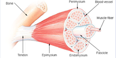
Skeletal Muscle Tissue Characteristics

answer
Muscle fibers ? force generation ?Multinucleated ?Nuclei at periphery Rich capillary network ?Helps tissue to meet metabolic demands Connective tissue ? force transmission ?Collagenous ?Fibers aligned parallel to greatest force
question
Development of Skeletal Muscle
answer
Develop from somites (dermomyotome) Myoblasts - migrate from somite ?Fuse to form myotube, which become myofibrils seen in adult tissue Fusion ? creates multinucleated cells (syncytium)
question
Cellular Characteristics of Skeletal Muscle

answer
Cytoplasm = sarcoplasm Cell membrane = sarcolemma ?Outer layer interacts w/ ECM and coatings ?Inner layer = true cell membrane Cellular contents ? primarily myofibrils, which provide appearance of muscle fibers
question
Cardiac Muscle
answer
Found only in heart Similar to skeletal muscle Continuously functioning Characteristics ?Net-like organization ? (more branching, less organization) ?Striated ?Complex junctions ?Intercalated disc - cellular junction b/w cells ?Fatigue resistance ?One or two nuclei ?Larger, more numerous T-tubules ?Extensive mitochondria
question
Net-like organization of cardiac muscle

answer
All of the tissue is really continuous w/ itself
question
Organization of the Myofibril
answer
Bands observable in light microscopy A band - large darkened central band I band - two light bands ?Split by Z-line H band - straddles M line Sarcomere extends from Z line to Z line
question
Sacromere
answer
Two filaments Thick ?Myosin (A band) Thin (Sacromere minus H zone) ?Actin ?Tropomyosin ?troponin M line ?Myosin anchoring site Z line ?Thin filament anchoring site
question
Thick Filament of Sarcomere

answer
Myosin - ATPase (co-factor - actin) ? one of the reasons that muscles require so much ATP ?Two heavy chains ?Four light chains Tail region (two heavy chains) Head region (globular ends of heavy chains plus light chains) ?Light chains wrapped around heavy chains Hinge region
question
Thin Filament of Sarcomere

answer
Composed of filamentous actin (F actin), tropomyosin and troponin Globular actin (G actin) binds myosin Tropomyosin bound to T actin Troponin bound to tropomyosin
question
Generation of Force in Skeletal Muscle
answer
Activation of contraction ?Influx of calcium into muscles ?Calcium binds troponin ?Troponin exposes actin ?Actin binds myosin ?Hinge ?Myosin moves against actin (power stroke) Contracted continues until calcium is depleted A single contraction may be result of hundreds actin myosin crossbridge building With contraction ?I band decreases in size ?H band decreases in size Actin-myosin crossbridge only released when myosin binds new ATP ?Rigor mortis Calcium comes from sarcoplasmic reticulum
question
Where does calcium come from?
answer
From the sarcoplasmic reticulum Triad: formed by sarcoplasmic reticulum ?Responsible for release of Ca to fibers, activation of muscle contraction Transverse (T) tubules ?Invaginations of sarcolemma ?Transmit depolarization ?Surround A and I bands Depolarization of cell membrane ?Continuous w/ T tubules ?Depolarization of T tubule physically stimulates SER ?SER releases Ca++ into cytoplasm Take home: T tubule network interacts w/ SER, which releases calcium for contraction
question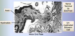
Myoneural Junction

answer
Motor nerves branch out w/in perimysial layer This interaction defines the function unit of muscle ? motor unit ?Defined as single neuron and the muscle fascicles it innervates ?If you want more precision in a movement, you need more neurons and therefore more motor units (i.e. hand)
question
Steps in Activation of Contraction
answer
?Steps in Activation of Contraction ?Action potential in Nerve Terminal ?Acetylcholine released (synaptic cleft) ?Binds receptors on sarcolemma ?Na+ influx ?Membrane depolarized ?Communicated via t tubules to SR ?Ca++ release from SR ?Contraction ?Excess acetylcholine hydrolyzed cholinesterases ?Muscle returns to resting state
question
Skeletal Muscle Transmission of Force
answer
Epimysium - External sheath of dense connective tissue (Entire muscle) Perimysium - Layer covering each bundle of muscle fibers (Muscle Fascicle) Endomysium - Layer covering each individual muscle fiber itself For the most part, the action that the muscle has will be transmitted to the bone or whatever else is moving as well
question
Dystroglycan Complex
answer
Dystrophin, dystroglycans (alpha and beta), sarcogylcans (alpha, beta, gamma and delta), and associated proteins (NOS, etc. Function: ?Connect actin to ECM (ECM makes up endomysium) ?Transmit generated force
question
Dystroglycan Complex Pathologies
answer
Duchenne Muscular Dystrophy - mutation in Dystrophin Limb Girdle muscular dystrophy (LGMD) - mutations in Dystroglycans, Sarcoglycans or other associated proteins
question
Transmission of Force in Cardiac Muscle
answer
Individual cells attached to each other by intercalated discs 3 components of intercalated disc are critical ?fascia adherens (hemi-Z bands) ? sacromeres to membrane ?same protein in Z band that attaches to thin filament ?macula adherens ? cell to cell ?gap junctions ? communications (allows for coordinated action of adjacent cells)
question
Smooth Muscle
answer
Two forms: single unit smooth muscle vs. multiunit smooth muscles Single Unit Smooth Muscle (single neuron, many cells) ?Found in walls of hollow viscera ?Function: propelling luminal contents, regulating flow of contents Multiunit Smooth Muscles (single neuron, single cell) ?i.e. trachea, iris of eye, hairs of skin ?Function: piloerection, shape changes
question
Tissue Organization of Smooth Muscle
answer
Interlaced bundles of muscle fibers allowing peristalsis Three-dimensionality
question
Generation of Force in Smooth Muscle
answer
Actin and myosin interaction (but on a cellular basis, not large macro basis like skeletal muscle) Lattice-like network Mediated by Ca++
question
Transmission of Force in Smooth Muscle
answer
Dense Bodies (attachment points) ?Two types: membrane and cytoplasmic ?Attachment for thin and intermediate filaments ?Composed of alpha-actinin (Z disc protein) ?Allow transmission of force from cell to cell
question
Muscle Tissue Function Overview
answer
Common Aspect of All Muscle Tissue Function - Contractibility Function ?Smooth Muscle - Individual Cell Basis ?Cardiac Muscle - Interconnected latticework of muscle fibers ?Skeletal Muscle - Functional unit is entire muscle Cross sectional area - Muscle Properties Sites of Attachment - Tendon Properties
question
Filament Polarity
answer
All of the actin molecules on one side of the Z line face a particular direction ?In the middle of the myosin filament, the heads change direction as well The importance of the polarity of the molecules is that actin and myosin can have directed interaction that ultimately causes sarcomere to shorten
question
Sarcomeric Shortening
answer
The filaments do not change length themselves They slide together to cause the structure as a whole to shorten
question
Sarcomere Length:Tension Relation
answer
Passive vs. Active ?Passive is what you get when muscle is not activated ?When you activate muscle, you get more force Force for active muscle is total force minus passive force After a certain point of stretch, active component of the force significantly decreases ?Thick and thin filaments are no longer able to overlap ? activation yields no force If the sarcomere is too short, thin filaments can actually interfere w/ each other, decreasing active component of force ?At some point, it gets so short that the thick filaments begin to crash into Z-line, which causes large resistance to contraction
question
Isometric Contraction
answer
Stimulating muscles but not allowing it to actually contract ? just measuring force generated upon activation
question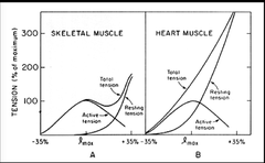
Tension Relation: Comparing Skeletal and Cardiac Muscle

answer
Cardiac muscle operates at a shorter sarcomere length (even though filaments are the same size) ?The more you fill the heart, the more you stretch out heart walls ?This causes sarcomeres to be able to create more active force ? this is a good thing ?We want heart to be able to contract more strongly
question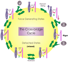
Cross-Bridge Cycling

answer
Rigor is state that will be reached when there is no ATP there or ADP greatly outnumbers ATP Power stroke of myosin moves filaments in sarcomere ?ATP comes in, there is some swinging of myosin head that occurs, myosin must attach and detach from actin, ultimately leads to actin filament being propelled with respect to the myosin filament ATP is split before myosin is activated ? only binds after activation step
question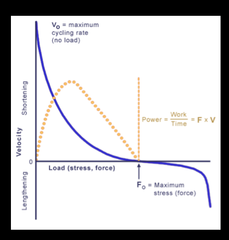
Force-Velocity Relationship

answer
The faster the muscle is allowed to shorten, the less force it can create There is a maximum rate that the muscle with shorten - Vo ?Limited by biochemical steps Maximum isometric force - Fo ?In order to lengthen muscle, you need to exceed that load
question
Excitation-Contraction Coupling Apparatus: Skeletal vs. Cardiac
answer
Cardiac muscle makes much less sarcotubule connections Due to different mechanisms of calcium release ?Cardiac muscle doesn't depend on actual physical junction, b/c the wave of calcium release can go along starting at one part and traveling along to the other
question
E-C Coupling in Cardiac Muscle: Calcium-induced calcium release
answer
Calcium from T-tubule binds to SR calcium release channel After a time, SR calcium release channel inactivates ?Important b/c it keeps cardiac muscle from being able to tetanize
question
Control of Contraction: Calcium binds to regulatory complex
answer
Binding site on actin is blocked by tropomyosin When calcium binds to troponin complex, tropomyosin moves and myosin can now attach to actin
question
Neural Control: Twitch
answer
Calcium reaches a peak and then starts trailing off While that is happening, myosins reach up and grab actin ?Takes some time for this to occur ? peak of twitch will occur some time after calcium peak
question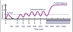
From Twitch to Tetanus

answer
Series of stimulations can lead to tetanus The closer together you place stimulation, the higher and higher the tension will remain, until the stimulations come rapidly enough together to maintain steady level of Ca in cell, which maintains steady level of tension ? tetanus
question
E-C Coupling in Smooth Muscle: Calcium entry into cytoplasm
answer
Membrane depolarization or first messenger binding to receptor can increase calcium levels within cell Increasing cytosolic calcium binds to calmodulin, which in turn binds to myosin-light chain kinase (MLCK) ?MLCK phosphorates myosin-light chain Start w/ inactive myosin ? activated by kinase ? binds to actin ?Turned off by phosphatase
question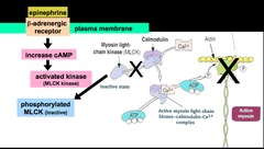
Smooth Muscle Relaxation via Epinephrine

answer
Effect of epinephrine depends on function of particular smooth muscle Epi binds to beta-adrenergic receptor, leads to increased cAMP cAMP activates MLCK kinase, which phosphorylates MLCK (now inactive) MLCK cannot phosphorylate (and therefore cannot activate) myosin, so it will not bind actin
question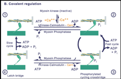
Smooth Muscle: Latch Bridge

answer
Can keep going over and over and over again, as long as myosin head keeps getting phosphorylated ? rapidly cycling phase If head is attached to actin, and the regulatory phosphate is pulled off, it creates a latch bridge ? holds on for dear life ?Eventually, it will let go, but it takes a very long time relative to rapidly cycling phase ?Maintain fixed tension against load Latch bridge is a good thing to have in vascular wall, to maintain position of vascular wall w/o requiring multiple ATPs to do so
question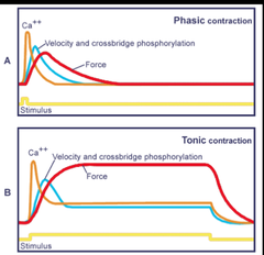
Smooth Muscle: Phasic vs. Tonic

answer
High level of force can be maintained w/ sustained stimulus
question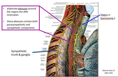
ANS is diffuse and difficult to dissect

answer
Will form a complex that looks like spiderwebs and will distribute itself everywhere Will latch onto spinal nerves
question
Somatic Motor vs. Visceral Motor

answer
Somatic: ?one neuron b/w CNS ; motor tissue ?cell body in CNS Visceral: ?Two neurons b/w CNS ; motor tissue ?Preganglionic neuron = cell body in CNS ?Postganglionic neuron = cell body in ganglion
question
Effect of Sympathetics vs. Parasympathetics
answer
Sympathetics = fight or flight (energy expenditure) ?Pupils dilate ?Bronchial dilation ?Vasoconstriction (skin) ?Quiet bowel ?sweating Parasympathetics = rest and digest (energy intake ; conservation) ?Pupils constrict ?Active bowel ?Relax anal and urinary sphincters "Fright" is a total ANS response ? both sympathetic and parasympathetics
question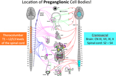
Location of Preganglionic Cell Bodies (Sympathetic vs. Parasympathetic)

answer
Preganglionic sympathetics = thoracolumbar (T1-L2/L3 of spinal cord) Preganglionic parasympathetics = craniosacral ?Brain: CN III, VII, IX, X ?Spinal cord: S2-S4
question
Location of Postganglionic Cell Bodies (Sympathetic vs. Parasympathetic)

answer
Sympathetics = all levels of spinal cord Parasympathetics = on or very lose to target organ/gland
question
Comparison of Length of Neurons
answer
Symp: short preganglionic, long postganglionic Parasymp: long preganglionic, short postganglionic ?i.e. vagus nerve
question
Visceral Motor Targets
answer
Symp: internal organs AND body wall Parasymp: internal organs ONLY
question
Specific Nerves of Parasympathetics
answer
Cranial Nerves ?Oculomotor (CN III) ? to ciliary ganglion (constrict pupil) ?Facial (CN VII) ? to pterygopalatine (stimulate lacrimal gland), submandibular ganglia (sensation to submandibular and lingual glands, travels w/ chorda tympani) ?Glossopharyngeal (CN IX) ? otic ganglion (parotid gland) ?Vagus (CN X) ? to ganglia in heart, larynx, trachea, bronchi, lungs, liver, gallbladder, stomach, pancreas, kidney, small intestine, and ascending colon Pelvic splanchnic nerves ?S2-S4 lateral horn
question
Sympathetics - Body Wall
answer
Visceral motor tissues: glands Smooth muscle: vascular smooth muscle, arrector pili muscles
question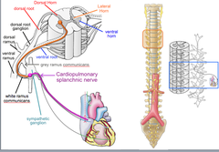
Postganglionic Cell Bodies of Sympathetics

answer
Paravertebral ganglia ? parallel to vertebral column ?Form a chain linked by preganglionic neuron that come from lateral horns from T1-L2/3 ?Therefore, some preganglionics must also ascend or descend in trunk to reach the cervical through sacral and coccygeal ganglia ?Postganglionic cell bodes in those cervical through coccygeal sympathetic ganglia ?Cardiopulmonary splanchnic nerves use the sympathetic ganglion to get to their target Prevertebral (preaortic) ganglia ?Anterior to abdominal aorta, near its major branches ?Named for major aortic branches ?Celiac ganglion ?Superior mesenteric ganglion ?Inferior mesenteric ganglion ?Preganglionic neurons reach prevertebral ganglia via abdominopelvic splanchnic nerves ?Instead of ascending or descending to reach target, they go through splanchnic nerve
question
Somatic vs. Visceral Sensory
answer
Somatic is restricted to skeletal muscle and connective tissue Visceral includes smooth muscle, cardiac muscle and glands Cell body outside of CNS ?Long peripheral process (dendrite), short central process (axon) ?Neural impulse: peripheral to central (afferent)
question
Functional Sensory Modalities: Somatic vs. Visceral
answer
Somatic Sensory ?Type: touch, temperature, cutting, ischemia ?Range: pleasant ; uncomfortable ; painful ?Region recognition: well-localized ?Pathway: somatic sensory fibers in spinal nerves from skin and skeletal muscle Visceral sensory ?Type: regulatory, ischemia, distention, cramping ?Range: uncomfortable - painful ?Region recognition: poorly localized, dull ?Pathway: pain mostly w/ sympathetics back to T1-L2 spinal cord segments from most internal organs ?Unconscious (reflexive, regulatory) w/ parasympathetics only from internal organs
question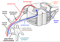
Pathway of Somatic Sensory

answer
Arrives from periphery Cell body in DRG Ultimately synapses in dorsal horn
question
Pathway of Visceral Sensory
answer
Returns to CNS via pathway of visceral motor (i.e. splanchnic nerves) No synapse in sympathetic ganglion Goes to spinal cord segments closest to white rami Ultimately synapse in dorsal horn ?Cross-talk can occur, so you can be tricked into thinking pain is coming from somewhere else (i.e. referred pain) ?Ex: diffuse chest /arm pain from heart injury
question
Musculoskeletal Pain
answer
E.g., patient describes a history of repetitive or unaccustomed activity involving the upper trunk or arms Often insidious and persistent Lasting for hours to weeks. Frequently sharp and localized to a specific area (such as the xiphoid, lower rib tips, or midsternum), but may be diffuse and poorly localized. The pain may be positional or exacerbated by deep breathing, turning, or arm movement
question
Non-ischemic cardiac chest pain
answer
May be caused by inflammation of the pericardium; inflammation of or injury to the myocardium w/o predisposing ischemic insult; or aortic injury. ?E.g., Pericarditis ?Typically of fairly sudden onset ?Occurs over the anterior chest ?Usually sharp and exacerbated by inspiration ?Pain may decrease in intensity when the patient sits up and can radiate, especially to the trapezius ridge
question
Visceral Referred Pain
answer
Information relayed to brain is segmental, not organ specific Brain "thinks" pain from a visceral structure is coming from an area of skin innervated by the same segmental level at which the visceral afferent synapses (in dorsal horn) Results from convergence of somatic and visceral afferents on the same segmental level of the spinal cord Signals transfer, which tricks the brain
question
Functions of Immune System
answer
Protection from invading substances Ability to distinguish "self"
question
Primary Lymphoid Organs
answer
Sites of immune cell production Bone marrow (B-cells), thymus (site of T-cell maturation)
question
Secondary Lymphoid Organs/Structures
answer
Organs ?Lymph nodes ?Spleen Structures ?MALT (mucosa associated lymphoid tissue) ?Includes solitary nodules, diffuse lymphatic tissue, structures grouped together like Peyer's patches ?Tonsils are stressed on exams
question
Immune System
answer
Cells ?Lymphocytes ?Accessory cells ?Effector cells Arrangement ?Loose ?Nodules ?MALT ?organs
question
Antigens
answer
Recognized by cells Epitopes - molecular domain that immune system mounts response to Humoral response = antibody removal of antigen Cellular response = lymphocyte removal of antigen
question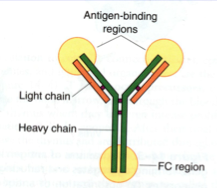
Antibodies

answer
Classes of immunoglobulins Glycoproteins ? allows for variety in side chains ?2 light chains, heavy chains Fc region and antigen binding site
question
How Antibodies Help Protect Organism
answer
Agglutination ?lymphocytes recognize foreign substance, they ramp up antibody production, antibodies attach to antigens, and when enough Abs bind it precipitates the antigen out ?neutralize harmful effects Phagocytosis (effect cells ? macrophages, neutrophils, eosinophils) ?Cells eat Opsonization and the Complement System ?Process of binding of antibodies to antigen, w/ complement proteins (produced in liver) recognized by effector cells and acting as extra receptors, combination of antibody/antigen becomes easier to recognize as foreign
question
Classes of Immunoglobins
answer
IgG - most abundant IgA - secreted IgM - activates B cells IgE - allergic reaction IgD - activates B cells
question
IgG
answer
Most abundant immunoglobulin (75-80%) IgGs cross the placenta - confer immunity to fetus during development Are activators of the complement system Act as opsonins
question
IgA
answer
Found in secretions 10-15% total serum immunoglobulins IgA secreting cells concentrate along mucous membranes IgA binding prevents pathogen attachment to cell
question
IgM
answer
First class of immunoglobulin produced in antigen response Membrane-bound on B cells Circulating form is a high valency pentamer Effective in agglutination and complement activation
question
IgE
answer
Low serum concentrations Mediate immediate-hypersensitivity reaction ?This is why we don't want too much of this immunoglobulin Receptors on mast cells and basophils ?IgE binds receptor and induces degranulation
question
IgD
answer
0.2% total serum immunoglobulins along w/ IgM, thought to activate B cells
question
Innate Immunity
answer
Innate, natural Anatomic barriers ?Intact skin ?Mucous membranes Physiologic barriers ?Temperature ?pH ?soluble factors endocytic and phagocytic barriers inflammatory response
question
Acquired Immunity
answer
Humoral first response Cell-mediated immunity Specific immunity Properties of Acquired Immunity: ?Specificity ?Diversity ?Memory ?Self-limitation ?Tolerance
question
Cells of the Immune System
answer
Lymphocytes - one epitope recognized All precursors are in the bone marrow ?B cells: memory and plasma cells ?T cells: memory and helper or cytotoxic Antigen presenting cells (APCs) ? accessory cells ?Dendritic cells (ex: langerhan's cells) ?macrophages Effector cells ?Cytotoxic T cells ?Helper T cells ?Plasma cells (focus on in histology) ?Mature B cells that have recognized antigen and are making antibodies
question
Lymphatic Tissue Structure
answer
Very cellular ? lots of cells that are multiplying and recognizing/removing antigens Scaffold of lymph node ? web of reticular fibers ?Made by reticular cells ?In thymus ? epithelial cell support (stellate) Rich supply of lymphocytes ?Morphologically indistinguishable, with exception of plasma cells
question
Lymphocytes
answer
Morphologically indistinguishable Plasma cells = only lympyocytes that are able to be differentiated ?"clock face" or "spokes of a wheel" ?Very granular and noticeable ?this is b/c they are very active
question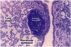
MALT

answer
Mucosa-associated lymphoid tissue Mostly IgA secreting cells, w/ APCs, B cells, helper T Cells ?APCs tend to be more towards germinal center Diffuse and solitary nodules Peyer's patches in ileum ?Collection of nodules under mucosa Tonsils - large collection of lymphoid nodules ?Palatine ?Pharyngeal ?lingual
question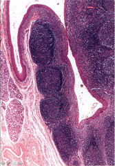
Histology of Tonsils

answer
IMAGE IS PALATINE TONSIL Palatine: ?Stratified squamous ?Crypts (infoldings ? while spaces in this image) Pharyngeal: ?Ciliated pseudostratified columnar (w/ goblet cells b/c it is a respiratory epithelium) ?No crypts Lingual: ?Stratified squamous ?One crypt (won't be on the test, b/c we would need to see the entire tonsil to make sure there is only one crypt)
question
Thymus
answer
Lymphoepithelial origin Cortex ?Thymocytes (T cell precursors ? mature here while filtering through) ?Epithelial reticular cells (actual support system of thymus) ?macrophages Medulla ?Differentiated T lymphocytes ?Epithelial reticular cells Thymic-blood barrier ? don't want precursor cells going out into blood until fully differentiated Histology ?Dark staining cortex, light staining medulla, incomplete trabeculae ?Hassall's corpuscles are diagnostic of thymus tissue ? thought to be degenerated epithelial reticular cells that have produced lots of concentric layers of keratin as they die
question
Lymph Node
answer
In-line lymph filter ?B cells and T cells monitor lymph for antigens here Cortex ?Inner ?Outer Medulla ?Cords ?sinuses Histology of Lymph Nodes ?Very defined dense irregular connective tissue capsule ?Spaces, open holes ? afferent lymph vessels ?Bring lymph to node, then it filters down through spaces ?Efferents collect lymph in medulla ?In the medulla, you can see plasma cells
question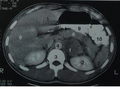
Spleen

answer
Largest lymphoid tissue Filter of blood ?Destruction of erythrocyte ?Blood circulation Capsule Hilum Splenic pulp ?White ?Red Histology ?Capsule - dense, irregular connective tissue that extends into parenchyma as incomplete trabeculae ?No subcapsular sinuses ?White pulp is not white on histological slide (named for what it looks like upon autopsy) ?Where response is mounted for immune system ?Red pulp = filtering of blood ?Splenic artery comes in through the hilum, will branch into trabecular arteries along w/ trabeculae, keeps branching until it gets to white pulp which has a central artery
question
White Pulp of Spleen
answer
Central artery is actually an arteriole ?Usually to the side of the lymphoid nodule ?Surrounded by smooth muscle ?Surrounded by sheath of T lymphocytes (periartieral lymphatic sheath = PALS) Lymphoid nodule is mostly B cells, w/ germinal center like a regular lymph nodule
question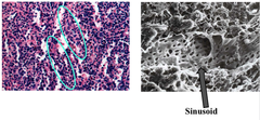
Red Pulp of Spleen

answer
Consists of splenic cords (of Billroth) and sinusoids ?Sinusoids are specialized capillaries w/ lots of spaces in it, allowing for bathing of cells in blood Circulation ?Closed = capillaries drain into sinusoids, and then back into capillary ?Open = arterial blood pass through cellular space and into sinusoids ?Return = sinusoid to red pulp vein to trabecular vein to splenic vein
question
Functions of Blood
answer
?Carry oxygen ?Remove waste ?Fight infection ?Bleeding/Stop bleeding ?Clotting/Stop clotting ?Carry nutrients
question
Hematopoiesis
answer
The formation of blood ?Granulopoiesis (granulocytes) ?Erythropoiesis (RBCs) ?Thrombopoiesis (platelets) ?Lymphopoiesis (lymphocytes) Pluripotent stem cell ? theoretical cell that has the ability to become any of the types of blood cells
question
Myeloid and Lymphoid Cells
answer
There are 4 primary categories of immune system ?Granulocytes (myeloid lineage) ?Lymphocytes (lymphoid lineage) ?Dendritic cells ?Lymphoid ?myeloid Some immune cells have multiple functions, leading to their classification under multiple cell categories
question
Hematopoietic Cell Lineage
answer
When a pluripotent stem cell divides, it forms two daughter cells that can differentiate into myeloid or lymphoid stem cells
question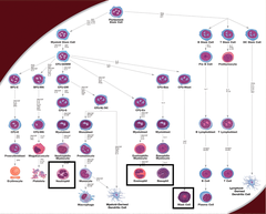
Granulocyte Lineage

answer
Granulocytes = cells that contain granules Includes neutrophils, eosinophils, basophils and mast cells
question
Lymphocyte Lineage
answer
Includes B cells and T cells
question
Erythrocyte Lineage
answer
RBCs are anuclear
question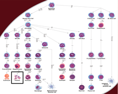
Platelet Lineage

answer
Derived from megakaryocytes Smallest cells
question
Monocyte Lineage
answer
Monocyte becomes macrophage involved in immune system
question
Mature Blood
answer
Contains: ?Plasma ? liquid portion of blood, has coagulation factors in it ?Serum = plasma w/o clotting factors ?Erythrocytes ?Leukocytes (WBCs) ?Neutrophils ?Basophils ?Eosinophils ?Monocyte/macrophages ?lymphocytes ?Platelets (thrombocytes)
question
Normal blood smears
answer
Mostly RBC Red outline w/ large nucleus = eosinophil White outline w/ large nucleus =neutrophil All blue stain = lymph Little dots = platelets
question
Granulopoiesis
answer
Myeloblast ? promyelocyte ? myelocyte ? metamyelocyte ?band cell ? segment (mature cell)
question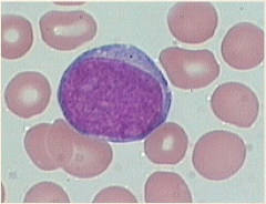
Myeloblast

answer
Most immature cell High nuclear:cytoplasmic ratio ? nucleus is majority of the cell Has nucleoli Large cell (the larger the white cell, the more immature it is) Hallmark of acute myelogenous leukemia (AML) ? it seen in peripheral smear, the patient has an acute leukemia
question
Promyelocyte
answer
Development of primary granules ?These granules are blue/purple High nuclear:cytoplasmic ratio Large cell ?Cell size progressive decreases w/ increasing differentiation/maturation Nucleoli present
question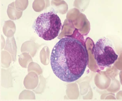
Myelocyte

answer
Cell starts to mature and you develop secondary granules (these will be present in adult cells) ?Red = eosinophil ?Purple = basophil ?Neutral = neutrophil Huff ? dawning of the myelocyte May have nucleoli This is the first stage that you can tell what the cell will be
question
Metamyelocyte

answer
Nuclear indentation occurs ?Must be less than 50% of the nucleus radius Continues to make secondary granules ?Primary granules essentially gone
question
Band cell

answer
aka stab When nuclear indentation becomes >50% Stage immediately before maturity
question
Action Potential
answer
Generated from initial segment of axon function of axon is to send info from cell body to other cells/neurons neurons are electrically polarized ? voltage difference across membrane = membrane potential ?b/c you have more negative charge inside of neurons than outside Membrane potential is maintained by ion channels and ion pumps ?Resting potential = -70mV ?If membrane potential rises high enough to initiate AP ? threshold potential = -55mV When stimulus arrives at cells, it will open up voltage gated Na+ channel, allowing Na+ influx ?Membrane potential rises from -70mV ?If it reaches -55mV, the AP will continue, if not, nothing happens ? all or none response When the threshold potential is reached, more voltage-gated Na+ channels open up, causing a more rapid Na+ influx ?Membrane potential becomes even more positive, reaching +30mV ?This is called the depolarization phase At +30mV, the membrane potential is reversed (now positive), inactivating the Na+ channels ?Potassium channels now open up, allowing potassium to flow from inside of cells to outside of cells ?This brings membrane potential back down towards resting potential ? repolarization phase Repolarization often has over shoot (as much as -90mV) ? hyperpolarization ?Protective mechanism ?Protects neurons from receives another stimulus ?At this point, sodium is actively pumped back out of the cell and the potassium is pumped back into the cell to regain the resting potential
question
Neutrophils

answer
Most prominent WBCs in blood (~75%) Main function: ?Eradicate pathogens ?Primary cell of innate immunity ?First to arrive in large numbers at site of infection (essentially, this is pus) Nucleus has 3-5 segments in an adult neutrophil
question
Eosinophils

answer
Grow up essentially the same as neutrophils, except that the granules are red ?Granules are also significantly larger Associations: ?Allergic rxn ?Helminthic infections Have capacity to express a variety of toxic oxidative intermediates Granules release: ?Major basic protein (MBP) ?Eosinophil peroxidase (EPO) ?Eosinophil cationic protein (ECP) ?Eosinophil-derived neurotoxin (EDN)
question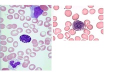
Basophils

answer
Rarest of white cells (usually <1%) ?Commonly found in myeloproliferative disease Found predominantly in blood but can migrate to tissue
question
Mast Cells
answer
Mature and are predominantly found in tissue
question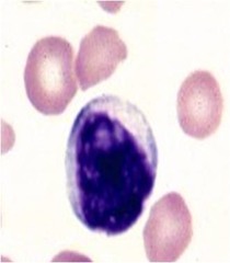
Lymphocyte

answer
Smaller than neutrophils Nucleus is most of the cell ?Scant cytoplasm ?Non-segmented nucleus Nucleus size is approximately the same size as an entire RBC Make the B & T cells
question
Plasma Cell

answer
Develop from B-lymphocyte Makes antibodies (immunoglobins) ?Each plasma cell only makes one class of Ig (IgM, IgA, IgD, IgG, IgE) Nucleus classically has clock-faced appearance, with slight huff next to nucleus
question
Monocytes
answer
The other non-segmented nuclear cell ?Larger than lymphocytes, more cytoplasm than lymphocytes ("flowing cytoplasm") Classified as phagocytes Have kidney bean nucleus Main functions: ?Phagocytosis ?Cell lysis
question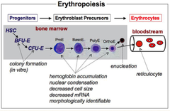
Erythropoiesis

answer
Occurs in bone marrow ?Only reticulocyte stage gets into blood ?Takes about 4 days to make new cell (3 days in marrow) ?The more you need new RBCs, the shorter the time in the bone marrow becomes Proerythroblast ? basophilic normoblast ? polychromatic normoblast ? orthochromic normoblast ? reticulocyte ? erythocyte RBC lineage is driven by erythropoietin ?Located on chromosome 7 ?Hormone secreted by kidney in response to renal hypoxia ?This causes secondary erythrocytosis (too many RBCs due in response to hypoxia)
question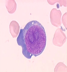
RBC begins as a proerythroblast

answer
Generally a large cell w/ nucleoli and round nucleus Stains blue
question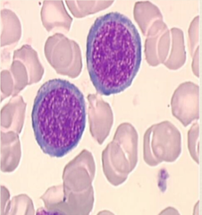
Basophilic normoblast

answer
Smaller than proerythroblast Chromatin gets tighter Cytoplasm becomes more blue
question
Polychromatophilic normoblast
answer
Cell gets even smaller, chromatin gets more condensed Cells have a multi-color appearance (cytoplasm blue-grey) b/c this is when the synthesis of hemoglobin starts ?Hemoglobin is red
question
Orthochromic Normoblast

answer
The chromatin material is very condensed now, the nucleus is small and the cytoplasm is very red (lots of hemoglobin)
question
Reticulocyte
answer
Nucleus has been extruded from cell Cytoplasm has a bluish hue (from leftover Has a slightly larger diameter than mature RBC Reticulocyte stain ? important b/c if someone is anemic to see if they are making new blood ?Low if they are not making new blood ?If someone IS making new blood and they are anemic, their reticular count should be high
question
Erythrocyte
answer
Biconcave disc Red b/c it is filled with hemoglobin Main function: ?Carries O2 ?Removes waste Has minor role in acid-base balance
question
Thrombocytopoiesis

answer
Megakaryocytes are very large cells found in bone marrow that will "bud" off and form platelets Platelets are the smallest cell in the peripheral blood (~2um) Platelets help to prevent bleeding ?Have granules that help to attract other cells and to prevent bleeding

